. 2006 Oct 3;45(39):12068-75.
doi: 10.1021/bi060899i.

Tropomodulin binds two tropomyosins: a novel model for actin filament capping

Affiliations

Tropomodulin binds two tropomyosins: a novel model for actin filament capping

Alla S Kostyukova et al. Biochemistry. .

Abstract

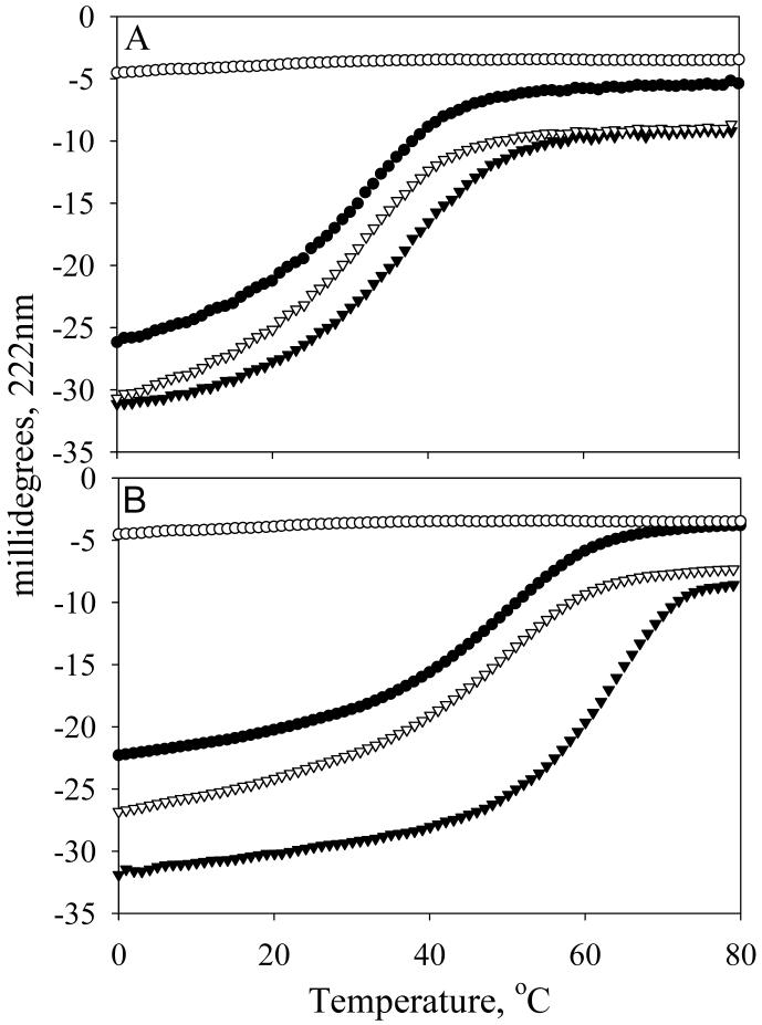
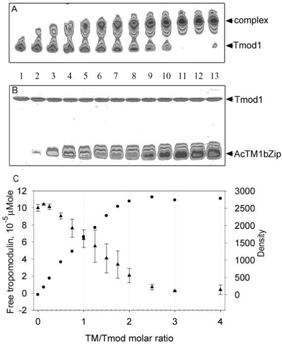
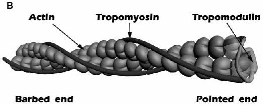
Tropomodulin, a tropomyosin-binding protein, caps the slow-growing (pointed) end of the actin filament regulating its dynamics. Tropomodulin, therefore, is important for determining cell morphology, cell movement, and muscle contraction. For the first time we show that one tropomodulin molecule simultaneously binds two tropomyosin molecules in a cooperative manner. On the basis of the tropomodulin solution structure and predicted secondary structure, we introduced a series of point mutations in regions important for tropomyosin binding and actin capping. Capping activity of these mutants was assayed by measuring actin polymerization using pyrene fluorescence. Using direct methods (circular dichroism and native gel electrophoresis) for detecting tropomodulin/tropomyosin binding, we localized the second tropomyosin-binding site to residues 109-144. Despite previous reports that the second binding site is for erythrocyte tropomyosin only, we found that both short nonmuscle and long muscle alpha-tropomyosins bind there as well, though with different affinities. We propose a model for actin capping where one tropomodulin molecule can bind to two tropomyosin molecules at the pointed end.


Figures

Figure 1
Figure 1
The amino acid sequence of Tmod1 aligned to the secondary structure obtained from NMR data (residues 1-38), secondary structure prediction (residues 39-159), crystal atomic structure (residues 160-344). Bold - helical regions where mutations were done. Underlined - residues that are mutated. Boxed residues represent fragments used for experiments.
Figure 2
Figure 2
Pointed end elongation of gelsolin-capped actin filaments (5.6 nM) in the presence and absence of tropomodulin and stTM. 1.1 μM (10 % pyrene-labeled) G-actin mixed with 0.6 μM F-actin in F-buffer (100 mM KCl, 2 mM MgCl2, 1 mM EGTA, 0.5 mM DTT, 0.2 mM ATP, 0.2 mM CaCl2, 1 mM NaN3, 10 mM imidazole, pH 7.0). (●) control, no tropomyosin or tropomodulin; (○) with 0.75 μM stTM; (▼) with 0.75 μM stTM and 20 nM Tmod1; (▽) with 0.75 μM stTM and 20 nM Tmod1(T59A/G60A/P61A); (■) with 0.75 μM stTM and 20 nM Tmod1(L27G); (□) with 0.75 μM stTM and 20 nM Tmod1(L27G/L71D).
Figure 3
Figure 3
Complex formation between AcTM1bZip and Tmod1 mutants monitored by non-denaturing polyacrylamide gel-electrophoresis. AcTM1bZip is positively charged and does not enter the gel. Lane 1 and lane 3, Tmod1; lane 2 and lane 4, Tmod1 and AcTM1bZip; lane 5, Tmod1(L27G); lane 6, Tmod1(L27G) and AcTM1bZip; lane 7, Tmod1(L27G/ L71D); lane 8, Tmod1(L27G/ L71D) and AcTM1bZip; lane 9, Tmod1(L27G/ L71D/I131D); lane 10, Tmod1(L27G/ L71D/I131D) and AcTM1bZip; lane 11, Tmod1(T59A/G60A/P61A); lane 12, Tmod1(T59A/G60A/P61A) and AcTM1bZip. TM:Tmod molar ratio in lane 4 is 1:1, in all other lanes with AcTM1bZip it is 2:1. Arrows indicate TM/Tmod1 complexes, arrowhead - Tmod1.
Figure 4
Figure 4
Binding of Tmod11-159 mutants to a TM model peptides measured using circular dichroism spectroscopy. The temperature dependence of the ellipticity at 222 nm was measured for unmixed and mixed AcTM1bZip and AcTM1aZip. A: Tmod11-159WT and AcTM1aZip; and B: Tmod11-159WT and AcTM1bZip; C: Tmod11-159(L27G/L71D) and AcTM1aZip; D: Tmod11-159(L27G/L71D) and AcTM1bZip. (○) Tmod11-159 alone; (●) TM peptide; (▽) sum of the folding curves of the TM peptide and Tmod11-159 alone; (▼) the folding of the mixture of the TM peptide and Tmod11-159.
Figure 5
Figure 5
Dependence of inhibition of actin polymerization on Tmod11-159 concentration in the presence of 0.75 μM stTM. Initial rates (R) were calculated as the first derivatives at time zero after fitting. The inhibition of polymerization was calculated as Rexp / Rcontrol, where Rcontrol=1 (in the absence of TM and Tmod1 fragments). (●) Tmod11-159WT; (○) Tmod11-159(L27G/L71D). (■) The value shown for stTM in the absence of tropomodulin fragments (n=3).
Figure 6
Figure 6
A: Pointed end elongation of gelsolin-capped actin filaments (5.6 nM) in the presence and absence of Tmod1(L27G/L71D/I131D) and 0.75 μM stTM. 1.1 μM (10 % pyrene-labeled) G-actin mixed with 0.6 μM F-actin in F-buffer (100 mM KCl, 2 mM MgCl2, 1 mM EGTA, 0.5 mM DTT, 0.2 mM ATP, 0.2 mM CaCl2, 1 mM NaN3, 10 mM imidazole, pH 7.0). (●) control, no tropomyosin or tropomodulin; (○) with 0.75 μM stTM and 50 nM Tmod1(L27G/L71D/I131D); (▼) with 0.75 μM stTM and 200 nM Tmod1 (L27G/L71D/I131D); (▽) with 0.75 μM stTM and 500 nM Tmod1(L27G/L71D/I131D); (■) with 500 nM Tmod1(L27G/L71D/I131D). B: Dependence of inhibition of actin polymerization on Tmod1 concentration. Initial rates (R) were calculated as the first derivatives at time zero after fitting. The inhibition of polymerization was calculated as Rexp / Rcontrol, where Rcontrol=1 (in the absence of TM and Tmod1 fragments). Tmod1WT with (●) and without (◆) 0.75 μM stTM; Tmod1(L27G/L71D) in presence of 0.75 μM stTM (○); Tmod1(L27G/L71D/I131D) with (▼) and without (◇) 0.75 μM stTM. (■) The value shown for stTM in the absence of tropomodulin (n=3).
Figure 7
Figure 7
Binding of Tmod1109-144 to TM model peptides measured using circular dichroism spectroscopy. The temperature dependence of the ellipticity at 222 nm was measured for unmixed and mixed AcTM1bZip and AcTM1aZip. A: Tmod1109-144 and AcTM1aZip; and B: Tmod1109-144 and AcTM1bZip. (○) Tmod1109-144 alone; (●) TM peptide; (▽) sum of the folding curves of the TM peptide and Tmod11-159 alone; (▼) the folding of the mixture of the TM peptide and Tmod1109-144.
Figure 8
Figure 8
Titration of Tmod1 by AcTM1bZip. Stock solution of Tmod1 was diluted to a final concentration of 10 μM with different concentrations of TM peptide. The decrease of free tropomodulin and the increase of the complex were monitored by scanning and quantifying the Tmod1 band in native polyacrylamide gels. A: 9 % native gel and B: 15 % SDS gel scanned on densitometer; C: dependence of amount of free Tmod1 (●) and density of the complex band (▲) on amount of AcTM1bZip added (TM/Tmod molar ratio). Lanes on the gels correspond to points on the graph. Lanes 1-13 contain AcTM1bzip/Tmod1 in ratios 0, 0.125, 0.25, 0.5, 0.75, 1.0, 1.25, 1.5, 1.75, 2.0, 2.5, 3.0, 4.0. Error bars show standard deviation (n=4).
Figure 9
Figure 9
A: schematic model of the TM-binding and actin-capping sites. B: a cartoon representation of the pointed end. The N-terminal half of Tmod1 binds to both TMs and interacts with actin. Two actin molecules at the pointed end are shown as transparent.
Figure 9
Figure 9
A: schematic model of the TM-binding and actin-capping sites. B: a cartoon representation of the pointed end. The N-terminal half of Tmod1 binds to both TMs and interacts with actin. Two actin molecules at the pointed end are shown as transparent.

References

    1. Pollard TD, Borisy GG. Cellular motility driven by assembly and disassembly of actin filaments. Cell. 2003;112:453–65. - PubMed
    1. Rafelski SM, Theriot JA. Crawling toward a unified model of cell mobility: spatial and temporal regulation of actin dynamics. Annu Rev Biochem. 2004;73:209–39. - PubMed
    1. Carlier MF, Pantaloni D. Control of actin dynamics in cell motility. J Mol Biol. 1997;269:459–67. - PubMed
    1. dos Remedios CG, Chhabra D, Kekic M, Dedova IV, Tsubakihara M, Berry DA, Nosworthy NJ. Actin binding proteins: regulation of cytoskeletal microfilaments. Physiol Rev. 2003;83:433–73. - PubMed
    1. Perry SV. Vertebrate tropomyosin: distribution, properties and function. J Muscle Res Cell Motil. 2001;22:5–49. - PubMed

Publication types