

doi: 10.1146/annurev.psych.58.110405.085709.
The experience of emotion
Affiliations
- PMID: 17002554
- PMCID: PMC1934613
- DOI: 10.1146/annurev.psych.58.110405.085709
Item in Clipboard
The experience of emotion
Annu Rev Psychol.
2007.
Abstract
Experiences of emotion are content-rich events that emerge at the level of psychological description, but must be causally constituted by neurobiological processes. This chapter outlines an emerging scientific agenda for understanding what these experiences feel like and how they arise. We review the available answers to what is felt (i.e., the content that makes up an experience of emotion) and how neurobiological processes instantiate these properties of experience. These answers are then integrated into a broad framework that describes, in psychological terms, how the experience of emotion emerges from more basic processes. We then discuss the role of such experiences in the economy of the mind and behavior.
Figures
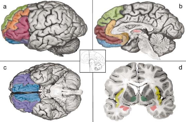
Key brain areas in the neural reference space for mental representations of emotion. The ventral system for core affect includes two closely connected circuits that are anchored in the orbitofrontal cortex (the entire ventral surface of the front part of the brain lying behind the orbital bone above the eye; Figure 1c). The sensory system involves the lateral sector of the orbitofrontal cortex (OFC) and includes the lateral portions of BA 11 and 13, BA 47/12 (a, c, purple). It is closely connected to the anterior insula (d, yellow) and the basolateral (BL) complex in the amygdala (d, rose, ventral aspect). The visceromotor circuitry includes the ventral portion of the ventromedial prefrontal cortex (VMPFC), which lies in the medial sector of the OFC (a, b, c, blue) and includes medial BA 11 and 13 ventral portions of BA 10, as well as BA 14, where the medial and lateral aspects of OFC connect; VMPFC is closely connected to the amygdala (including the central nucleus, d, rose, dorsal aspect) and the subgenual parts of the anterior cingulate cortex involving the anterior aspects of BA 24, 25, and 32 on the medial wall of the brain (ACC; b, copper and tan). The dorsal system is associated with mental state attributions including the dorsal aspect of the VMPFC corresponding to the frontal pole in BA 10 (b, maroon), the anterior ACC (peach), and the dorsomedial prefrontal cortex (DMPFC) corresponding to the medial aspects of BA 8, 9, and 10 (a, b, green). Ventrolateral prefrontal cortex (VLPFC) is shown in red (a). Also shown for reference are the thalamus (b, light pink), the ventral striatum (d, green), and the middle frontal gyrus in the dorsolateral prefrontal cortex (a, orange). Photographs adapted from DeArmond et al. (1989, pp. 5, 7, 8, and 43).
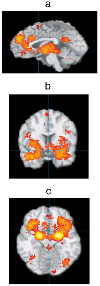
Preliminary summary of neuroimaging studies of core affective and emotion experiences. Activation foci were registered to a common stereotaxic brain atlas (Talairach & Tournoux 1988) where x = distance in millimeters to the right (+) or left (−) of midline; y = distance anterior (+) or posterior (−) to the anterior commissure; and z = distance superior (+) or inferior (−) to a horizontal plane through the anterior and posterior commissures. Midsagital (a, x = 0), coronal (b, y = 7), and horizontal (c, z = −13) images are presented. Significant areas of activation include OFC, insula, amygdala, ACC, and DMPFC (as well as VLPFC; not shown). VMPFC activations were also observed, but it is not clear that they extend down to the ventral surface, c, probably owing to problems with imaging that area of the brain. Lighter colors indicate a larger number of studies reported significant peak activations at that location (summary corrected for false discovery rate).
References
-
- Adolphs R. The neurobiology of social cognition. Curr. Opin. Neurobiol. 2001;11:231–39. - PubMed
-
- Adolphs R, Lee GP, Tranel D, Damasio AR. Bilateral damage to the human amygdala early in life impairs knowledge of emotional arousal. Soc. Neurosci. Abstr. 1997;23:1582.
-
- Allman JM, Hakeem A, Erwin JM, Nimchinsky E, Hoff P. The anterior cingulate cortex: the evolution of an interface between emotion and cognition. Ann. NY Acad. Sci. 2001;935:107–17. - PubMed
-
- Amaral DG, Behniea H, Kelly JL. Topographical organization of projections from the amygdala to the visual cortex in the Macaque monkey. Neuroscience. 2003;118:1099–120. - PubMed
-
- Anand A, Li Y, Wang Y, Wu J, Gao S, et al. Activity and connectivity of brain mood regulating circuit in depression: a functional magnetic resonance study. Biol. Psychiatry. 2005;57:1079–88. - PubMed
Publication types
MeSH terms
Grants and funding
LinkOut - more resources
Full Text Sources
