. 2006 Sep 26:4:32.
doi: 10.1186/1741-7007-4-32.

Modulation of Abeta42 low-n oligomerization using a novel yeast reporter system

Affiliations

Modulation of Abeta42 low-n oligomerization using a novel yeast reporter system

Sviatoslav Bagriantsev et al. BMC Biol. .

Abstract

Background: While traditional models of Alzheimer's disease focused on large fibrillar deposits of the Abeta42 amyloid peptide in the brain, recent work suggests that the major pathogenic effects may be attributed to SDS-stable oligomers of Abeta42. These Abeta42 oligomers represent a rational target for therapeutic intervention, yet factors governing their assembly are poorly understood.

Results: We describe a new yeast model system focused on the initial stages of Abeta42 oligomerization. We show that the activity of a fusion of Abeta42 to a reporter protein is compromised in yeast by the formation of SDS-stable low-n oligomers. These oligomers are reminiscent of the low-n oligomers formed by the Abeta42 peptide in vitro, in mammalian cell culture, and in the human brain. Point mutations previously shown to inhibit Abeta42 aggregation in vitro, were made in the Abeta42 portion of the fusion protein. These mutations both inhibited oligomerization and restored activity to the fusion protein. Using this model system, we found that oligomerization of the fusion protein is stimulated by millimolar concentrations of the yeast prion curing agent guanidine. Surprisingly, deletion of the chaperone Hsp104 (a known target for guanidine) inhibited oligomerization of the fusion protein. Furthermore, we demonstrate that Hsp104 interacts with the Abeta42-fusion protein and appears to protect it from disaggregation and degradation.

Conclusion: Previous models of Alzheimer's disease focused on unravelling compounds that inhibit fibrillization of Abeta42, i.e. the last step of Abeta42 assembly. However, inhibition of fibrillization may lead to the accumulation of toxic oligomers of Abeta42. The model described here can be used to search for and test proteinacious or chemical compounds for their ability to interfere with the initial steps of Abeta42 oligomerization. Our findings suggest that yeast contain guanidine-sensitive factor(s) that reduce the amount of low-n oligomers of Abeta42. As many yeast proteins have human homologs, identification of these factors may help to uncover homologous proteins that affect Abeta42 oligomerization in mammals.


Figures
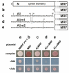
Figure 1
Figure 1
AβMRF causes nonsense suppression in yeast. (Upper panel) Schematic illustration of the constructs used in this study: (a) full length Sup35p (NMRF) (b) Sup35p without the N-terminal prion domain (MRF) (c) 42 fused to the N terminus of MRF (AβMRF) (d) AβMRF carrying a double mutation of Phe19,20Thr in its Aβ42 portion (Aβm1MRF) (e) AβMRF carrying a triple mutation of Phe19,20Thr and Ile31Pro in its Aβ42 portion (Aβm2MRF). All these constructs carry an HA tag between the M and RF domains. Images shown are not to scale. (Lower panel) Equal numbers of ade1-14 cells containing a genomic deletion of SUP35 (sup35Δ), and carrying the indicated constructs (a-e) on a plasmid were grown on complex medium, or synthetic medium supplemented (+Ade) or not (-Ade) with adenine. (a) Cells with inactivated NMRF ([PSI+]) had an impaired translational termination activity, were white and grew on -Ade. (b) Cells with fully active MRF (lacking the aggregation-prone prion, N, domain), were red and failed to grow on -Ade. (c) Cells expressing AβMRF have an impaired translational termination activity, as they were white and grew on -Ade. (d, e) The translational termination activity was restored by F19,20T (Aβm1MRF) and F19,20T/I31P (Aβm2MRF) mutations in the Aβ42 region of the fusion protein, making the cells dark pink and preventing their growth on -Ade.
Figure 2
Figure 2
AβMRF forms SDS-stable oligomers in yeast. (A) Immunoblot analysis of lysates from sup35Δ cells containing prionized NMRF ([PSI+]) or other indicated constructs. Lysates were treated with 1% SDS for 7 mins at room temperature and resolved by electrophoresis in agarose. Immunoblot analysis was performed using anti-RF antibodies, followed by stripping and staining with anti-Aβ antibodies. The positions of molecular weight standards, treated identically to the experimental samples, are shown (calc., calculated position). AβMRF formed SDS-stable low-n oligomers that largely disappeared after the introduction of the F19,20T (Aβm1MRF) and F19,20T/I31P (Aβm2MRF) mutations into the Aβ42 portion of the fusion protein. The decreased efficacy with which anti-Aβ antibodies recognized oligomers of AβMRF suggests that oligomerization occurred through the Aβ42 portion of the fusion protein. (B) 5 mg of amyloid fibers of Aβ42 peptide were treated with 1% SDS, resolved in agarose and analyzed by immunoblotting with anti-Aβ antibodies. Only a fraction of Aβ42 fibres can enter the 1.5% agarose gel. (C) Same as in (A) but the samples were resolved in an acrylamide gel. Asterisk denotes non-specific antibody interaction.
Figure 3
Figure 3
Guanidine stimulates oligomerization of AβMRF. sup35Δ cells expressing the indicated constructs were grown in the absence (-) or presence (+) of 6.3 mM guanidine (Gu). Equal amounts of lysate proteins were treated with 1% SDS and analyzed by immunoblotting with anti-RF or anti-Aβ antibodies following electrophoresis in agarose. Equal protein loading on each panel was confirmed by coomassie staining of the membrane (not shown).
Figure 4
Figure 4
Deletion of HSP104 decreases the total amount of AβMRF and reduces the proportion of oligomers. (A) AβMRF or Aβm2MRF were expressed in a sup35Δ strain in the presence (WT) or absence (Δ) of HSP104. Equal amounts of lysate proteins were treated with 1% SDS and analyzed by immunoblotting with anti-RF antibodies following electrophoresis in agarose. Equal protein loading was confirmed by coomassie staining of the membrane (not shown). (B) The effects of HSP104 deletion on the total amount of AβMRF and the ratio between oligomers and monomers from panel A were evaluated by densitometry. The height of the bars reflects total amount of AβMRF relative to that in HSP104 WT cells (error bars: s.e., n = 3). Each bar is subdivided according to the content of oligomers (open) and monomers (shaded) of AβMRF (± s.e., n = 3). The deletion of HSP104 decreased the total amount of AβMRF and decreased the ratio of oligomers to monomers.
Figure 5
Figure 5
Deletion of HSP104 exacerbates the translation termination defect of AβMRF. Equal numbers of sup35Δ yeast containing (WT) or lacking (Δ) HSP104 and expressing AβMRF or Aβm2MRF were grown on complex medium, or synthetic medium supplemented (+Ade) or not (-Ade) with adenine. Deletion of HSP104 stimulated growth of AβMRF-expressing cells on -Ade, while having no effect on yeast grown on +Ade medium.
Figure 6
Figure 6
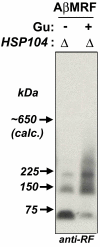
Guanidine stimulates oligomerization of AβMRF in the absence of HSP104. AβMRF-expressing sup35Δ hsp104Δ cells were grown in the absence (-) or presence (+) of 6.3 mM guanidine (Gu). Equal amounts of lysate proteins were treated with 1% SDS and analyzed by immunoblotting with anti-RF antibodies following electrophoresis in agarose. Equal protein loading was confirmed by coomassie staining of the membrane (not shown).
Figure 7
Figure 7
Co-immunoprecipitation of Hsp104 with AβMRF. Lysates of sup35Δ cells with (WT) or without (Δ) HSP104, expressing non-tagged NMRF, HA-tagged MRF, or HA-tagged AβMRF, were incubated with anti-HA antibodies immobilized on agarose beads. Co-precipitated proteins were eluted and analyzed by immunoblotting with anti-RF and anti-Hsp104 antibodies. Hsp104 co-immunoprecipitated with AβMRF, but not with MRF. Non-HA-tagged NMRF was used as a control for non-specific binding to anti-HA antibodies.

Similar articles

Cited by

References

    1. LaFerla FM, Oddo S. Alzheimer's disease: Abeta, tau and synaptic dysfunction. Trends Mol Med. 2005;11:170–176. doi: 10.1016/j.molmed.2005.02.009. - DOI - PubMed
    1. Lesne S, Koh MT, Kotilinek L, Kayed R, Glabe CG, Yang A, Gallagher M, Ashe KH. A specific amyloid-beta protein assembly in the brain impairs memory. Nature. 2006;440:352–357. doi: 10.1038/nature04533. - DOI - PubMed
    1. Hardy J, Selkoe DJ. The amyloid hypothesis of Alzheimer's disease: progress and problems on the road to therapeutics. Science. 2002;297:353–356. doi: 10.1126/science.1072994. - DOI - PubMed
    1. Walsh DM, Selkoe DJ. Deciphering the molecular basis of memory failure in Alzheimer's disease. Neuron. 2004;44:181–193. doi: 10.1016/j.neuron.2004.09.010. - DOI - PubMed
    1. Klein WL, Stine WB, Jr, Teplow DB. Small assemblies of unmodified amyloid beta-protein are the proximate neurotoxin in Alzheimer's disease. Neurobiol Aging. 2004;25:569–580. doi: 10.1016/j.neurobiolaging.2004.02.010. - DOI - PubMed

Publication types

MeSH terms