. 2006 Sep 26:4:35.
doi: 10.1186/1476-7120-4-35.

Non-compacted cardiomyopathy: clinical-echocardiographic study

Affiliations

Non-compacted cardiomyopathy: clinical-echocardiographic study

Nilda Espinola-Zavaleta et al. Cardiovasc Ultrasound. .

Abstract

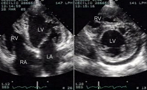
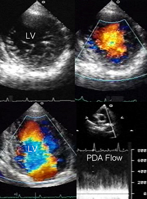
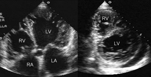
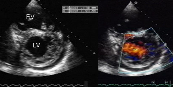
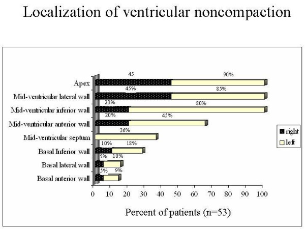
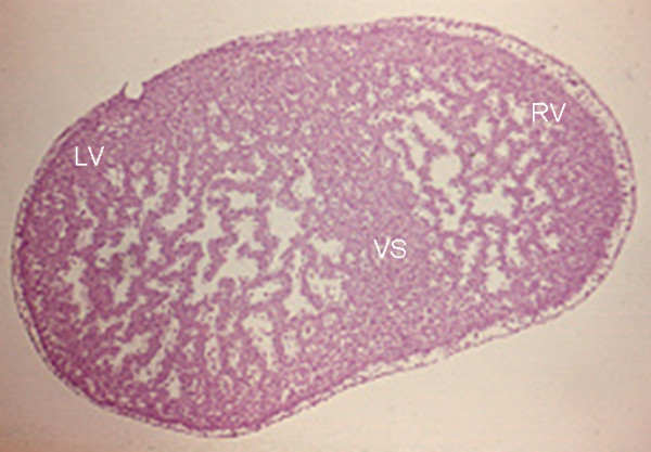
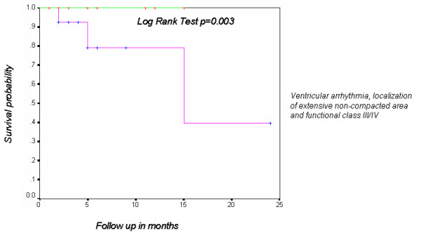
The aim of the present study was to describe the clinical and echocardiographic findings of ventricular noncompaction in adult patients. Fifty-three patients underwent complete clinical history, electrocardiogram, Holter and transthoracic echocardiogram. Forty patients (75%) were in class I/II of the New York Heart Association, and 13 (25%) in class III/IV. Ventricular and supraventricular escape beats were found in 40% and 26.4%, respectively. Holter showed premature ventricular contractions in 32% and sustained ventricular tachycardia in 7.5%. Ventricular noncompaction was an isolated finding in 74% of cases and was associated with other congenital heart disease in 26%. Noncompacted ventricular myocardium involved only left ventricle in 62% of the patients and both ventricles in 38%. The mean ratio of noncompacted to compacted myocardial layers at the site of maximal wall thickness was 3.4 +/- 0.87 mm (range 2.2-7.5). The presence of ventricular noncompaction in more than three segments was associated with a functional class greater than II and ventricular arrhythmia with demonstrable statistical significance by chi2(p < 0.003).

Conclusion: a) Noncompacted cardiomyopathy is a congenital pathological entity that can occur in isolated form or associated with other heart disease and often involves both ventricles. b) A ratio of noncompacted to compacted myocardium greater than 3 and involvement of three or more segments are indicators of poor prognosis. c) Since the clinical manifestations are not sufficient to establish diagnosis, echocardiography is the diagnostic tool that makes it possible to document ventricular noncompaction and establish prognostic factors.


Figures

Figure 1
Figure 1
Two-dimensional apical four chamber and parasternal short axis images at the level of the ventricles show dilatation of both ventricles, multiple trabeculae and intertrabecular recesses in inferior, lateral, anterior walls, middle and apical portions of the septum and apex of the left ventricle. A mild pericardial effusion can be observed. LV: Left ventricle; LA: Left atrium; RV: Right ventricle; RA: Right atrium.
Figure 2
Figure 2
Transthoracic two-dimensional study with color and continuous wave Doppler shows left ventricular noncompaction associated with patent ductus arteriosus (PDA). Trabeculae and deep recesses with penetration of color can be observed in the left ventricle. Continuous wave Doppler from a suprasternal approach at the level of the great vessels registers systolic-diastolic flow through the ductus arteriosus. Others abbreviations as before.
Figure 3
Figure 3
Transthoracic two-dimensional echocardiogram in apical four chamber and parasternal short axis at the level of both ventricles demonstrate dilatation, deep trabeculae and intertrabecular recesses in the inferior, lateral, anterior walls, middle and apical portions of the septum and apex of the left ventricle. The right ventricle also shows evidence of noncompaction. A posterolateral pericardial effusion is also present. Others abbreviations as before.
Figure 4
Figure 4
Two-dimensional parasternal and color Doppler images at the level of both ventricles that show the noncompacted:compacted wall ratio and how the color enters the intertrabecular recesses. Others abbreviations as before.
Figure 5
Figure 5
Graph shows the percentage of different segments of the left and right ventricular wall affected by noncompaction.
Figure 6
Figure 6
Microphotograph of a transverse section at the level of both ventricles of a heart that shows extensively developed trabeculae that fill the ventricular lumen. Note the form of the more compacted ventricular septum (arrow). 250 X. VS-Ventricular septum. Others abbreviations as before.
Figure 7
Figure 7
Kaplan Meier curve that shows the probability of survival in patients with ventricular arrhythmia, localization of extensive non-compacted area and functional class III/IV.

Similar articles

Cited by

References

    1. Maron BJ, Towbin JA, Thiene G, Antzelevich C, Corrado D, Arnett D, Moss AJ, Seidman CE, Young JB. AHA Scientific Statement. Contemporary Definition and Classification of the Cardiomyopathies. Circulation. 2006;113:1807–1816. doi: 10.1161/CIRCULATIONAHA.106.174287. - DOI - PubMed
    1. Thiene G, Corrado D, Basso C. Cardiomyopathies: Is it time for a molecular classification? Eur Heart J. 2004;25:1772–1775. doi: 10.1016/j.ehj.2004.07.026. - DOI - PubMed
    1. Ritter M, Oechslin E, Sutsch G, Attenhofer C, Schnider J, Jenni R. Isolated noncompaction of the myocardium in adults. Mayo Clin Proc. 1997;72:26–31. - PubMed
    1. Weiford BC, Subbarao VD, Mulhern KM. Noncompaction of the ventricular myocardium. Circulation. 2004;109:2965–2971. doi: 10.1161/01.CIR.0000132478.60674.D0. - DOI - PubMed
    1. Dusek J, Ostadal B, Duskova M. Postnatal persistence of spongy myocardium with embryonic blood supply. Arch Pathol. 1975;99:312–317. - PubMed