

A hypothesis for vulnerable plaque rupture due to stress-induced debonding around cellular microcalcifications in thin fibrous caps
- PMID: 17003118
- PMCID: PMC1595411
- DOI: 10.1073/pnas.0606310103
A hypothesis for vulnerable plaque rupture due to stress-induced debonding around cellular microcalcifications in thin fibrous caps
Abstract
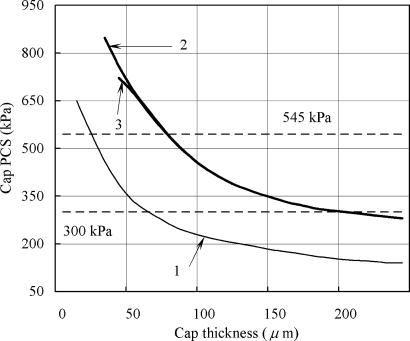
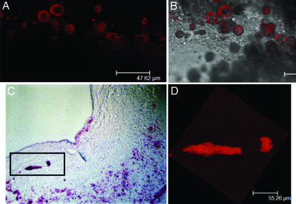
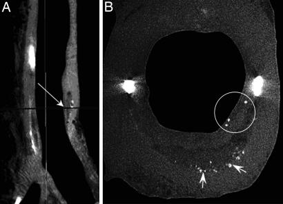
In this article, we advance a hypothesis for the rupture of thin fibrous cap atheroma, namely that minute (10-mum-diameter) cellular-level microcalcifications in the cap, which heretofore have gone undetected because they lie below the visibility of current in vivo imaging techniques, cause local stress concentrations that lead to interfacial debonding. New theoretical solutions are presented for the local stress concentration around these minute spherical inclusions that predict a nearly 2-fold increase in interfacial stress that is relatively insensitive to the location of the hypothesized microinclusions in the cap. To experimentally confirm the existence of the hypothesized cellular-level microcalcifications, we examined autopsy specimens of coronary atheromatous lesions using in vitro imaging techniques whose resolution far exceeds conventional magnetic resonance imaging, intravascular ultrasound, and optical coherence tomography approaches. These high-resolution imaging modalities, which include confocal microscopy with calcium-specific staining and micro-computed tomography imaging, provide images of cellular-level calcifications within the cap proper. As anticipated, the minute inclusions in the cap are very rare compared with the numerous calcified macrophages observed in the necrotic core. Our mathematical model predicts that inclusions located in an area of high circumferential stress (>300 kPa) in the cap can intensify this stress to nearly 600 kPa when the cap thickness is <65 microm. The most likely candidates for the inclusions are either calcified macrophages or smooth muscle cells that have undergone apoptosis.
Conflict of interest statement
The authors declare no conflict of interest.
Figures
Similar articles
-
Micro-CT based analysis of a new paradigm for vulnerable plaque rupture: cellular microcalcifications in fibrous caps.Mol Cell Biomech. 2008 Mar;5(1):37-47. Mol Cell Biomech. 2008. PMID: 18524245
-
Size and proximity of micro-scale hard-inclusions increase the risk of rupture in fibroatheroma-like laboratory models.J Mech Behav Biomed Mater. 2023 May;141:105749. doi: 10.1016/j.jmbbm.2023.105749. Epub 2023 Mar 6. J Mech Behav Biomed Mater. 2023. PMID: 36924613 Free PMC article.
-
Microcalcifications, Their Genesis, Growth, and Biomechanical Stability in Fibrous Cap Rupture.Adv Exp Med Biol. 2018;1097:129-155. doi: 10.1007/978-3-319-96445-4_7. Adv Exp Med Biol. 2018. PMID: 30315543 Review.
-
Influence of microcalcifications on vulnerable plaque mechanics using FSI modeling.J Biomech. 2008;41(5):1111-8. doi: 10.1016/j.jbiomech.2007.11.029. Epub 2008 Feb 7. J Biomech. 2008. PMID: 18258240
-
Calcium deposition within coronary atherosclerotic lesion: Implications for plaque stability.Atherosclerosis. 2020 Aug;306:85-95. doi: 10.1016/j.atherosclerosis.2020.05.017. Epub 2020 Jun 14. Atherosclerosis. 2020. PMID: 32654790 Review.
Cited by
-
Volumetric quantification of fibrous caps using intravascular optical coherence tomography.Biomed Opt Express. 2012 Jun 1;3(6):1413-26. doi: 10.1364/BOE.3.001413. Epub 2012 May 16. Biomed Opt Express. 2012. PMID: 22741086 Free PMC article.
-
Detection of hydroxyapatite in calcified cardiovascular tissues.Atherosclerosis. 2012 Oct;224(2):340-7. doi: 10.1016/j.atherosclerosis.2012.07.023. Epub 2012 Jul 24. Atherosclerosis. 2012. PMID: 22877867 Free PMC article.
-
Intracranial Arterial Calcifications: Potential Biomarkers of Stroke Risk and Outcome.Front Neurol. 2022 Sep 1;13:900579. doi: 10.3389/fneur.2022.900579. eCollection 2022. Front Neurol. 2022. PMID: 36119671 Free PMC article. Review.
-
Inhibition of bone morphogenetic protein signal transduction prevents the medial vascular calcification associated with matrix Gla protein deficiency.PLoS One. 2015 Jan 20;10(1):e0117098. doi: 10.1371/journal.pone.0117098. eCollection 2015. PLoS One. 2015. PMID: 25603410 Free PMC article.
-
Effect of calcification on the mechanical stability of plaque based on a three-dimensional carotid bifurcation model.BMC Cardiovasc Disord. 2012 Feb 15;12:7. doi: 10.1186/1471-2261-12-7. BMC Cardiovasc Disord. 2012. PMID: 22336469 Free PMC article.
References
-
- Falk E. Circulation. 1992;86(Suppl 3):30–42. - PubMed
-
- Lendon CL, Davies MJ, Born GV, Richardson PD. Atherosclerosis. 1991;87:87–90. - PubMed
-
- Libby P. Circulation. 1995;91:2844–2850. - PubMed
-
- Little WC. Am J Cardiol. 1990;66:44G–47G. - PubMed
-
- Muller JE, Tofler GH. Ann Epidemiol. 1992;2:393–405. - PubMed
MeSH terms
LinkOut - more resources
Full Text Sources
Other Literature Sources
Medical
Research Materials
Miscellaneous
