. 2006 Oct 3;103(40):14672-7.
doi: 10.1073/pnas.0602952103. Epub 2006 Sep 26.

Templated biomineralization on self-assembled protein fibers

Affiliations

Templated biomineralization on self-assembled protein fibers

K Subburaman et al. Proc Natl Acad Sci U S A. .

Abstract

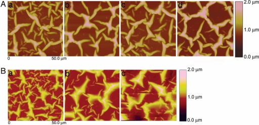
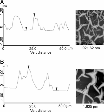
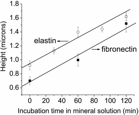
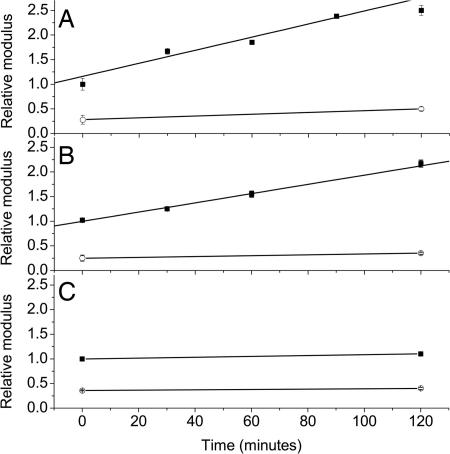
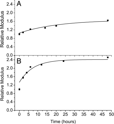
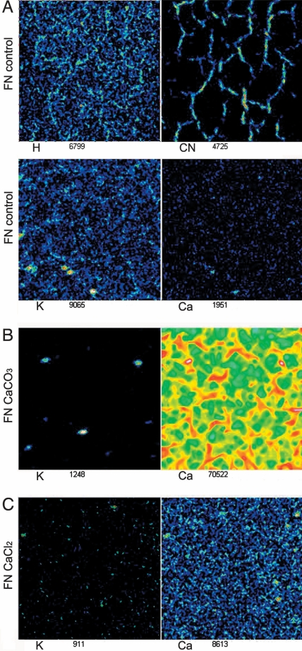
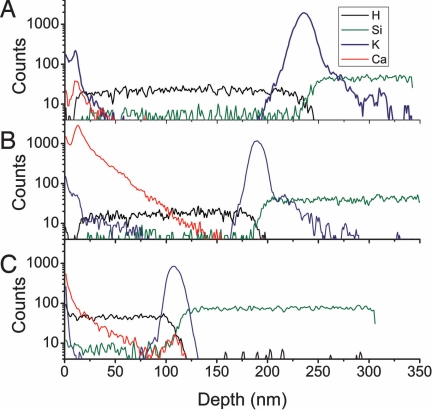
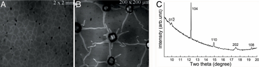
Biological mineralization of tissues in living organisms relies on proteins that preferentially nucleate minerals and control their growth. This process is often referred to as "templating," but this term has become generic, denoting various proposed mineral-organic interactions including both chemical and structural affinities. Here, we present an approach using self-assembled networks of elastin and fibronectin fibers, similar to the extracellular matrix. When induced onto negatively charged sulfonated polystyrene surfaces, these proteins form fiber networks of approximately 10-mum spacing, leaving open regions of disorganized protein between them. We introduce an atomic force microscopy-based technique to measure the elastic modulus of both structured and disorganized protein before and during calcium carbonate mineralization. Mineral-induced thickening and stiffening of the protein fibers during early stages of mineralization is clearly demonstrated, well before discrete mineral crystals are large enough to image by atomic force microscopy. Calcium carbonate stiffens the protein fibers selectively without affecting the regions between them, emphasizing interactions between the mineral and the organized protein fibers. Late-stage observations by optical microscopy and secondary ion mass spectroscopy reveal that Ca is concentrated along the protein fibers and that crystals form preferentially on the fiber crossings. We demonstrate that organized versus unstructured proteins can be assembled mere nanometers apart and probed in identical environments, where mineralization is proved to require the structural organization imposed by fibrillogenesis of the extracellular matrix.


Conflict of interest statement

The authors declare no conflict of interest.

Figures

Fig. 1.
Fig. 1.
Schematic cross-section of the ECM fiber network self-assembled upon a thin protein layer on spin-coated SPS. Relative scales of AFM tip and fiber network are indicated.
Fig. 2.
Fig. 2.
AFM images (50 × 50 μm) showing network of proteins after exposure to CaCO3 mineralization conditions for different times. (A) Elastin at 0 min (a), 60 min (b), 90 min (c), and 120 min (d). (B) Fibronectin at 0 min (a), 60 min (b), and 120 min (c). (Magnifications: ×800.)
Fig. 3.
Fig. 3.
Height profiles obtained by AFM along indicated lines of the elastin network after immersion in calcium bicarbonate solution for 0 min (A) and 120 min (B). (Magnifications: ×600.)
Fig. 4.
Fig. 4.
Average height of elastin and fibronectin fibers imaged by AFM as a function of immersion time with the free drift mineralization method.
Fig. 5.
Fig. 5.
Relative modulus of elastin (A) and fibronectin (B) fibers (■) and the flat regions (○) measured by using SMFM as a function of immersion time with the free drift mineralization method, and elastin fiber in CaCl2 solution (C).
Fig. 6.
Fig. 6.
Relative modulus of elastin (A) and fibronectin (B) fibers measured by using SMFM as a function of immersion time with the flow cell mineralization method.
Fig. 7.
Fig. 7.
TOF-SIMS chemical maps of fibronectin (A) and fibronectin mineralized by using the flow cell method for 48 h (B) and fibronectin immersed in CaCl2 sample for 24 h (C). Note that Ca is drastically enhanced on the fibers in the mineralized sample. (Magnifications: ×800.)
Fig. 8.
Fig. 8.
TOF-SIMS depth profile of fibronectin (A), fibronectin mineralized with flow cell method (B), and fibronectin immersed in a CaCl2 sample (C) for H, Si, K, and Ca. Depth has been calibrated by depth crater.
Fig. 9.
Fig. 9.
Late mineralization data. (A and B) Optical micrographs showing crystal formation on elastin fibers after 24 h mineralization by the free drift method, indicating presence of crystals on the fiber vertices. (Magnifications: A, ×20; B, ×200.) (C) Synchrotron x-ray diffraction pattern from the same conditions (λ = 0.6525 Å).

References

    1. Schinke T, McKee MD, Karsenty G. Nat Genet. 1999;21:150–151. - PubMed
    1. Hunter GK, Goldberg HA. Proc Natl Acad Sci USA. 1993;90:8562–8565. - PMC - PubMed
    1. Murshed M, Shinke T, McKee MD, Karsenty G. J Cell Biol. 2004;165:625–630. - PMC - PubMed
    1. Singla A, Lee CH. J Biomed Mater Res. 2002;60:368–374. - PubMed
    1. Service RF. Science. 1999;286:2442–2444.

Publication types

LinkOut - more resources