. 2006 Oct 3;103(40):14813-8.
doi: 10.1073/pnas.0602678103. Epub 2006 Sep 26.

Transaldolase is essential for maintenance of the mitochondrial transmembrane potential and fertility of spermatozoa

Affiliations

Transaldolase is essential for maintenance of the mitochondrial transmembrane potential and fertility of spermatozoa

Andras Perl et al. Proc Natl Acad Sci U S A. .

Abstract

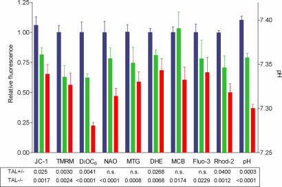
Fertility of spermatozoa depends on maintenance of the mitochondrial transmembrane potential (Deltapsi(m)), which is generated by the electron-transport chain and regulated by an oxidation-reduction equilibrium of reactive oxygen intermediates, pyridine nucleotides, and glutathione (GSH). Here, we report that male mice lacking transaldolase (TAL)(-/-) are sterile because of defective forward motility. TAL(-/-) spermatozoa show loss of Deltapsi(m) and mitochondrial membrane integrity because of diminished NADPH, NADH, and GSH. Mitochondria constitute major Ca(2+) stores; thus, diminished mitochondrial mass accounts for reduced Ca(2+) fluxing, defective forward motility, and infertility. Reduced forward progression of TAL-deficient spermatozoa is associated with diminished mitochondrial reactive oxygen intermediate production and Ca(2+) levels, intracellular acidosis, and compensatory down-regulation of carbonic anhydrase IV and overexpression of CD38 and gamma-glutamyl transferase. Microarray analyses of gene expression in the testis, caput, and cauda epididymidis of TAL(+/+), TAL(+/-), and TAL(-/-) littermates confirmed a dominant impact of TAL deficiency on late stages of sperm-cell development, affecting the electron-transport chain and GSH metabolism. Stimulation of de novo GSH synthesis by oral N-acetyl-cysteine normalized the low fertility rate of TAL(+/-) males without affecting the sterility of TAL(-/-) males. Whereas TAL(-/-) sperm failed to fertilize TAL(+/+) oocytes in vitro, sterility of TAL(-/-) sperm was circumvented by intracytoplasmic sperm injection, indicating that TAL deficiency influenced the structure and function of mitochondria without compromising the nucleus and DNA integrity. Collectively, these data reveal an essential role of TAL in sperm-cell mitochondrial function and, thus, male fertility.


Conflict of interest statement

The authors declare no conflict of interest.

Figures

Fig. 1.
Fig. 1.
Male infertility in TAL-deficient mice. Fifteen male and 15 female mice of each genotype were paired, with a single WT female or male per cage. Values indicate mean number ± SD of pups per breeding pair recorded for four generations. Based on comparison with TAL+/+ controls, P values <0.05 are shown.
Fig. 2.
Fig. 2.
Cumulative assessment of Δψm, mitochondrial mass, ROI, GSH, and cytoplasmic and mitochondrial Ca2+ levels in live spermatozoa from TAL+/+, TAL+/− and TAL−/− littermates. Results were expressed as relative fluorescence values with respect to those of TAL+/+ cells normalized at 1.0 on the left y axis. Absolute pH values based on SNARF-1 fluorescence of calibration standards are shown on the right y axis. Data present mean ± SE of eight independent experiments. P values under each column reflect comparison of TAL+/− (green) and TAL−/− (red) spermatozoa to TAL+/+ (blue) ones. n.s., not significant.
Fig. 3.
Fig. 3.
Loss of Δψm and structural changes in mitochondria of TAL-deficient sperm. (A) Fluorescence microscopy of spermatozoa from TAL+/+, TAL+/−, and TAL−/− mice. Cells were stained with DiOC6 (green) and DAPI (blue) to detect Δψm of mitochondria in the midsection and DNA in heads of sperm cells, respectively. BF, bright-field images. With respect to TAL+/+ sperm, Δψm was progressively diminished in TAL+/− and TAL−/− sperm. (Magnification, ×400.) (B) Transmission EM of cauda and caput epididymidis and testis from TAL+/+ and TAL−/− littermate mice. Original magnification is shown above images of cauda epididymidis. Mitochondria (M) are arranged in a cylinder-shaped sheath that wraps spirally around the axial outer dense fiber–axoneme complex of the midpiece (MP). No structural defects were observed in the head and tail section of TAL−/− spermatozoa. The head (H) was of normal size and shape. The axoneme complex (AC) of TAL−/− sperm showed the expected pattern of two central microtubules, surrounded by nine tubules, intact dynein arms, and a set of outer dense fibers. Although the outer mitochondrial membranes were appropriately retained, the internal membrane structure of mitochondria was irregular, with disappearance of cristae-like folds (white arrows) and formation of electron-dense central clumps in mitochondria of TAL−/− spermatozoa in cauda and caput epididymidis (black arrows).
Fig. 4.
Fig. 4.
HPLC analysis of PPP sugars and nucleotides. (A) HPLC analysis of S7P in testis of TAL+/+, TAL+/−, and TAL−/− mice. Chromatograms indicate electric charge (nanocoulomb) of sugar phosphates recorded on a pulse amperometric detector. S7P was positively identified by spiking TAL+/+ tissue extract with sugar phosphate standard (data not shown). Bar charts show comparative analysis of S7P levels in testis from age-matched TAL+/+, TAL+/−, and TAL−/− littermates. Data show mean ± SE of measurements from four mice per genotype. P values <0.05 reflect differences between genotypes. (B) Analysis of nucleotides in cauda epididymidis of TAL+/+, TAL+/−, and TAL−/− mice by HPLC. Chromatograms indicate UV absorbance (AU). Annotated peaks were positively identified by spiking TAL+/+ tissue extracts separately with each compound (data not shown). Bar charts show comparative analysis of nucleotide levels in tissues from age-matched TAL+/+, TAL+/−, and TAL−/− littermates. Data show mean ± SE of measurements from four mice per genotype. P values <0.05 are indicated.
Fig. 5.
Fig. 5.
Effect of TAL deficiency on expression of CAIV, CD38, and GGT. (A) Western blot analysis of CAIV expression in testis and caput and cauda epididymidis of TAL+/+, TAL+/−, and TAL−/− littermates. Blots were first developed with rabbit antibody to CAIV then reexposed to antibodies to TAL and actin. By using automated densitometry, CAIV expression levels in each tissue were normalized to actin levels and compared with TAL+/+ cells set at 1.0. (B) Expression of CD38 on the surface of sperm cells from TAL+/+, TAL+/−, and TAL−/− littermates gated on live cells based on forward scatter and side scatter. Antibody to CD59 was used as a control. Histograms of TAL+/− and TAL−/− spermatozoa (shaded curves) are overlayed on those from TAL+/+ littermate controls (open curves). Data represent analysis of five experiments. (C) Activity of TAL, TK, G6PD (left y axis), and GGT (right y axis) in cauda epididymidis of TAL+/+, TAL+/−, and TAL−/− mice. Data represent mean ± SD of four sets of TAL+/+, TAL+/−, and TAL−/− littermates. ∗, P < 0.05; ∗∗, P < 0.01; ∗∗∗, P < 0.0001.

Similar articles

Cited by

References

    1. Fisher HM, Aitken RJ. J Exp Zool. 1997;277:390–400. - PubMed
    1. Marchetti C, Obert G, Deffosez A, Formstecher P, Marchetti P. Hum Reprod. 2002;17:1257–1265. - PubMed
    1. Marchetti C, Jouy N, Leroy-Martin B, Defossez A, Formstecher P, Marchetti P. Hum Reprod. 2004;19:2267–2276. - PubMed
    1. Perl A, Gergely P, Jr, Puskas F, Banki K. Antiox Redox Signal. 2002;4:427–443. - PubMed
    1. Mayes PA. In: Harper's Biochemistry. Murray RK, Granner DK, Mayes PA, Rodwell VW, editors. Norwalk, CT: Appleton & Lange; 1993. pp. 201–211.

Publication types

MeSH terms

Associated data