

Interferon-gamma plays protective roles in sodium arsenite-induced renal injury by up-regulating intrarenal multidrug resistance-associated protein 1 expression
- PMID: 17003472
- PMCID: PMC1780179
- DOI: 10.2353/ajpath.2006.060024
Interferon-gamma plays protective roles in sodium arsenite-induced renal injury by up-regulating intrarenal multidrug resistance-associated protein 1 expression
Abstract
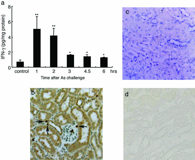
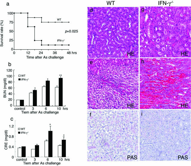
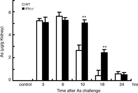
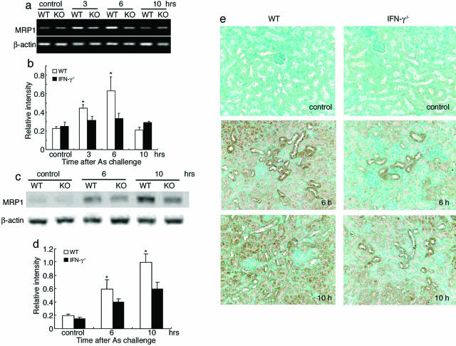
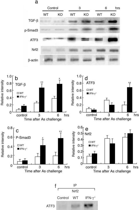
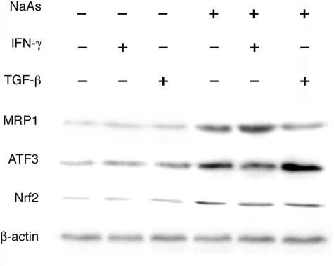
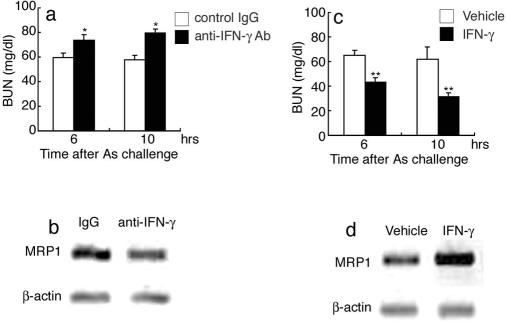
Subcutaneous injection of sodium arsenite (NaAs, 12.5 mg/kg) into BALB/c [wild-type (WT)] mice causes acute renal dysfunction characterized by severe hemorrhages, acute tubular necrosis, and cast formation, with increases in serum blood urea nitrogen and creatinine levels. Concomitant enhancement in intrarenal interferon (IFN)-gamma expression prompted us to examine its roles in this pathology. IFN-gamma-deficient (IFN-gamma-/-) mice exhibited higher serum blood urea nitrogen and creatinine levels and exaggerated histopathological changes, compared with WT mice. Eventually, IFN-gamma-/- mice exhibited a high mortality (87.5%) within 24 hours after NaAs challenge, whereas most WT mice survived. The intrarenal arsenic concentration was significantly higher in IFN-gamma-/- mice later than 10 hours after NaAs treatment, with attenuated intrarenal expression of multidrug resistance-associated protein (MRP) 1, a main transporter for NaAs efflux, compared with WT mice. NF-E2-related factor (Nrf) 2 protein, a transcription factor crucial for MRP1 gene expression, was similarly increased in the kidneys of both strains of mice after NaAs treatment. In contrast, the absence of IFN-gamma augmented transforming growth factor-beta-Smad3 signal pathway and eventually enhanced the expression of activating transcription factor 3, which is presumed to repress Nrf2-mediated MRP1 gene expression. Thus, IFN-gamma can protect against NaAs-induced acute renal injury, probably by maintaining Nrf2-mediated intrarenal MRP1 gene expression.
Figures
Similar articles
-
MRP-1 expression levels determine strain-specific susceptibility to sodium arsenic-induced renal injury between C57BL/6 and BALB/c mice.Toxicol Appl Pharmacol. 2005 Feb 15;203(1):53-61. doi: 10.1016/j.taap.2004.07.013. Toxicol Appl Pharmacol. 2005. PMID: 15694464
-
The absence of interleukin-6 enhanced arsenite-induced renal injury by promoting autophagy of tubular epithelial cells with aberrant extracellular signal-regulated kinase activation.Am J Pathol. 2010 Jan;176(1):40-50. doi: 10.2353/ajpath.2010.090146. Epub 2009 Dec 11. Am J Pathol. 2010. PMID: 20008137 Free PMC article.
-
Interferon-γ is protective in cisplatin-induced renal injury by enhancing autophagic flux.Kidney Int. 2012 Nov;82(10):1093-104. doi: 10.1038/ki.2012.240. Epub 2012 Jul 11. Kidney Int. 2012. PMID: 22785177
-
IFN-gamma protects cerulein-induced acute pancreatitis by repressing NF-kappa B activation.J Immunol. 2007 Jun 1;178(11):7385-94. doi: 10.4049/jimmunol.178.11.7385. J Immunol. 2007. PMID: 17513789
-
Rifampicin-induced injury in HepG2 cells is alleviated by TUDCA via increasing bile acid transporters expression and enhancing the Nrf2-mediated adaptive response.Free Radic Biol Med. 2017 Nov;112:24-35. doi: 10.1016/j.freeradbiomed.2017.07.003. Epub 2017 Jul 6. Free Radic Biol Med. 2017. PMID: 28688954
Cited by
-
Mechanisms pertaining to arsenic toxicity.Toxicol Int. 2011 Jul;18(2):87-93. doi: 10.4103/0971-6580.84258. Toxicol Int. 2011. PMID: 21976811 Free PMC article.
-
Decreased cohesin in the brain leads to defective synapse development and anxiety-related behavior.J Exp Med. 2017 May 1;214(5):1431-1452. doi: 10.1084/jem.20161517. Epub 2017 Apr 13. J Exp Med. 2017. PMID: 28408410 Free PMC article.
-
TrxR1 as a potent regulator of the Nrf2-Keap1 response system.Antioxid Redox Signal. 2015 Oct 1;23(10):823-53. doi: 10.1089/ars.2015.6378. Epub 2015 Jun 24. Antioxid Redox Signal. 2015. PMID: 26058897 Free PMC article. Review.
-
Nrf2 protects human bladder urothelial cells from arsenite and monomethylarsonous acid toxicity.Toxicol Appl Pharmacol. 2007 Dec 1;225(2):206-13. doi: 10.1016/j.taap.2007.07.016. Epub 2007 Aug 7. Toxicol Appl Pharmacol. 2007. PMID: 17765279 Free PMC article.
-
STAT1 regulates interferon-γ-induced angiotensinogen and MCP-1 expression in a bidirectional manner in primary cultured mesangial cells.J Renin Angiotensin Aldosterone Syst. 2020 Jul-Sep;21(3):1470320320946527. doi: 10.1177/1470320320946527. J Renin Angiotensin Aldosterone Syst. 2020. PMID: 32741247 Free PMC article.
References
-
- Goering PL, Aposhian HV, Mass MJ, Cebrian M, Beck BD, Waalkes MP. The enigma of arsenic carcinogenesis: role of metabolism. Toxicol Sci. 1999;49:5–14. - PubMed
-
- Klaassen CD. Heavy metals and heavy-metal antagonist. Hardman JG, Gilman AG, Limbird LE, editors. New York: McGraw-Hill,; Goodman and Gilman’s The Pharmacological Basis of Therapeutics, (ed 9) 1996:pp 1649–1672.
-
- Liu J, Liu YP, Goyer RA, Chanzar W, Waalkes MP. Metallothionein-I/II null mice are more sensitive than wild-type mice to the hepatotoxic and nephrotoxic effects of oral or injected inorganic arsenicals. Toxicol Sci. 2000;55:460–467. - PubMed
-
- Snow E. Metal carcinogenesis: mechanistic implications. Pharmacol Ther. 1992;53:31–65. - PubMed
-
- Thompson DJ. A chemical hypothesis for arsenic methylation in mammals. Chem Biol Interact. 1993;88:89–114. - PubMed
MeSH terms
Substances
LinkOut - more resources
Full Text Sources
Other Literature Sources
Molecular Biology Databases
Research Materials
Miscellaneous
