. 2006 Oct;169(4):1155-66.
doi: 10.2353/ajpath.2006.060058.

alpha-1 antitrypsin inhibits caspase-3 activity, preventing lung endothelial cell apoptosis

Affiliations

alpha-1 antitrypsin inhibits caspase-3 activity, preventing lung endothelial cell apoptosis

Irina Petrache et al. Am J Pathol. 2006 Oct.

Abstract

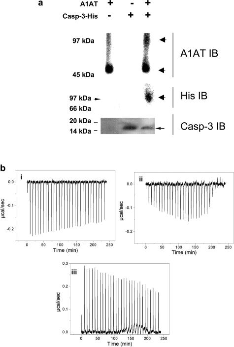
alpha-1 Antitrypsin (A1AT) is an abundant circulating serpin with a postulated function in the lung of potently inhibiting neutrophil-derived proteases. Emphysema attributable to A1AT deficiency led to the concept that a protease/anti-protease imbalance mediates cigarette smoke-induced emphysema. We hypothesized that A1AT has other pathobiological relevant functions in addition to elastase inhibition. We demonstrate a direct prosurvival effect of A1AT through inhibition of lung alveolar endothelial cell apoptosis. Primary pulmonary endothelial cells internalized human A1AT, which co-localized with and inhibited staurosporine-induced caspase-3 activation. In cell-free studies, native A1AT, but not conformers lacking an intact reactive center loop, inhibited the interaction of recombinant active caspase-3 with its specific substrate. Furthermore, overexpression of human A1AT via replication-deficient adeno-associated virus markedly attenuated alveolar wall destruction and oxidative stress caused by caspase-3 instillation in a mouse model of apoptosis-dependent emphysema. Our findings suggest that direct inhibition of active caspase-3 by A1AT may represent a novel anti-apoptotic mechanism relevant to disease processes characterized by excessive structural cell apoptosis, oxidative stress, and inflammation, such as pulmonary emphysema.


Figures

FIGURE 1
FIGURE 1
Antiapoptotic effects of hA1AT in lung microvascular endothelial cells. a: Caspase-3 activity (fold increase versus vehicle-treated control) in mouse lung endothelial cells exposed to staurosporine (ST, 600 nmol/L; 2 hours) in the presence of purified hA1AT (0.05 mg/ml, 16 hours; mean + SEM, n = 3, P < 0.05 versus ctl* or versus ST#). b: Cytochrome c release (normalized by protein concentration) induced by staurosporine in the presence of increasing concentrations of A1AT (pretreatment for 16 hours; mean + SEM, n = 2, P < 0.05 versus ctl* or ST#). c: DAPI-stained nuclei in blue in mouse lung endothelial cells exposed to staurosporine (ST, 600 nmol/L; 2 hours) in the presence of purified hA1AT [0.05 mg/ml, 16 hours, or vehicle (Veh)]. Note a reduction of total number of attached cells, which now show small nuclear size with pyknotic morphology in the ST-treated cells (arrows), and in contrast, a preserved number of nuclei of normal morphology in cells pretreated with hA1AT. Representative experiment, n = 4. Scale bar = 10 μm.
FIGURE 2
FIGURE 2
a: Immunoblots of nuclear (n) and cytoplasmic (c) fractions of mouse cells obtained after incubation with purified hA1AT (0.05 mg/ml, 24 hours) in the presence or absence of fetal bovine serum in the media, followed by washing and removal of the media. Note the hA1AT uptake in the cytoplasmic fraction, confirmed by the presence of actin immunostaining. Importantly, there was no cross-reactivity with the endogenous mouse or the serum bovine A1AT. n = 1 experiment. b–e: Fluorescence micrographs of mouse cells after incubation with Dylight alone (b) or with Dylight-labeled h1AT (0.05 mg/ml, 4 hours, c). In d, cells were counterstained with DAPI in blue to visualize nuclei 2 hours after the addition of hA1AT. Note hA1AT uptake in the cytoplasm (arrow) (in b–e, assays were performed in nonpermeabilized, live cells, representative of n = 3 experiments). e: Confocal microscopy of mouse endothelial cells treated with Dylight-hA1AT (0.05 mg/ml, 16 hours) confirming the presence of hA1AT intracellular (cytoplasm, arrow; nucleus, arrowhead; n = 2 experiments).
FIGURE 3
FIGURE 3
hA1AT interaction with active caspase-3 in cultured lung endothelial cells. a: Co-immunofluorescence of hA1AT and caspase-3 in mouse and rat microvascular lung endothelial cells. i and ii: hA1AT (green) co-localizing (yellow, arrow) with active caspase-3 (red) in mouse endothelial cells. iii: hA1AT (red) co-localizing (yellow, arrow) with active caspase-3 (green) in apoptotic cells (staurosporine-treated, 600 nmol/L, 2 hours; n = 2 experiments), but not with procaspase-3 (iv, arrowhead; untreated) or with rabbit IgG (v, arrowhead; untreated). b: Co-immunoprecipitation studies in mouse cells (treated with ST, 600 nmol/L, 2 hours; n = 2) with antibodies against caspase-3 for immunoprecipitation followed by immunoblotting for hA1AT before (top) and after (middle) incubation with A1AT blocking peptide. After stripping and reprobing, immunoprecipitated caspase-3 was confirmed by blotting for active caspase-3 (bottom). Lane 1: Starting material: diluted total cell lysate. Lane 2: Immunoprecipitate with active caspase-3-specific monoclonal antibody. Lane 3: Immunoprecipitate with a polyclonal antibody specific for active and procaspase-3. Lane 4: hA1AT protein control. Note that both caspase-3 antibodies precipitated hA1AT (lanes 2 and 3, top) and the lack of hA1AT detection in the presence of a specific hA1AT-blocking peptide.
FIGURE 4
FIGURE 4
hA1AT interaction with active caspase-3 in cell-free systems. a: Native gel electrophoresis of the complex of hA1AT with histidine (His)-tagged active caspase-3. Top: immunoblot with hA1AT antibody highlighting the presence of an estimated 97-kd complex revealing the presence of His-caspase-3, after stripping and reprobing with His antibody (middle); (because of the nondenaturing conditions of migration, the molecular weight of the markers may not be accurate). The SDS-PAGE (bottom) reveals His-tagged active caspase-3 by Western blot, representative of two experiments). b: Isothermal calorimetry of the hA1AT-caspase-3 interaction. Exothermic enthalpies measured on hA1AT (0.098 mmol/L/injection) incubation with i) active caspase-3 peptide (0.0089 mmol/L, n = 1), ii) elastase (0.0097 mmol/L, n = 3), as positive control, or iii) PBS, as negative control (n = 3). Note the incubation with buffer generated an endothermic interaction.
FIGURE 5
FIGURE 5
Functional effects of hA1AT on caspase-3 activity in cell-free systems. a: Dose-dependent inhibitory effect of hA1AT on the enzymatic activity (expressed in fluorescence signal per unit of time) of recombinant active caspase-3 on the specific substrate DEVD (P < 0.05). b: Kinetic curves of caspase-3 activity (cleavage of DEVD substrate) measured after incubating recombinant human active caspase-3 with vehicle (black triangles) or with bovine serum albumin (gray circles, 120 mg/ml, BSA), α-2 macroglobulin (red squares; 60 mg/ml, A2MG), or increasing concentrations of hA1AT. Inset showing the effects on caspase-3 activity for two lower concentrations of A1AT (100 and 500 nmol/L) compared with vehicle (Veh). c: Effect of hA1AT on caspase-3 activity (percent inhibition of maximum) compared with α1-antichymotrypsin (ACT), α2-macroglobulin (α2MG), or serum albumin (BSA) (mean, n = 2). d: Inhibitory effect of hA1AT on the enzymatic activity of recombinant active caspase-3 versus purified elastase (percent inhibition of maximum activity, representative of n = 20). In the inset we reported data as ratios of caspase-3 activity (fluorescence units) when incubated with A1AT versus the activity of uninhibited caspase-3, representing the reduction factors for A1AT.
FIGURE 6
FIGURE 6
Effect of posttranslational modifications of A1AT on its anti-caspase-3 activity. a: Effect of polymerization (poly A1AT; 60°C for 2 hours) and oxidation, obtained by exposure to cigarette smoke extract (Oxy−), on the caspase-3 inhibitory effect of hA1AT (percent inhibition of maximum activity) compared with native hA1AT (n = 5, *P < 0.05). b: Effects A1AT conformers on caspase-3 activity [activity of uninhibited caspase-3 in buffer is noted as vehicle (3) and is superimposed on the RCL-cleaved poly A1AT (2) curve]. The reactive center loop (RCL)-cleaved A1AT (via Staphylococcus aureus protease V8) and the polymerized RCL-cleaved A1AT (1) (via papain) (n = 2) were tested against controls that included the above proteases (5) and against polymerized noncleaved hA1AT, Poly(M) (4). The right inset depicts the concentration-dependence of the inhibitory activity against caspase-3 for the (M) polymers and for the RCL-cleaved polymers of A1AT. c: Effect of the cleaved hA1AT peptide, the C-36 fibrillary fragment, on caspase-3 activity (mean + SEM, n = 3; *P < 0.05).
FIGURE 7
FIGURE 7
Protective effect of hA1AT on alveolar space destruction and oxidative stress triggered by lung instillation of active caspase-3 in mice. a: Morphometric measurements of alveolar perimeters, and b: mean linear intercept 48 hours after intratracheal caspase-3 or Chariot vehicle (Ctl) administration in the presence of hA1AT-expressing adeno-associated virus (hA1AT −AAV) or control virus (c-AAV) given by intramuscular injection (mean + SD, P < 0.05 versus c-AAV* and versus hA1AT-AAV+ caspase-3, analysis of variance, posthoc t-test, n = 4 to 5 mice per group). c: Histology of H&E-stained lung alveolar tissue in mice receiving intratracheal chariot vehicle (Ctl) or caspase-3 in the presence of control virus (c-AAV), or hA1AT-expressing adeno-associated virus (hA1AT −AAV). Note the airspace enlargement in the mice receiving active caspase-3 and empty vehicle (arrows), which is attenuated in the presence of overexpressed hA1AT. d: Lung catalase activity 2 days after intratracheal instillation of active caspase-3 or Ctl (mean + SEM, *P < 0.05 versus ctl #P < 0.05 versus caspase-3). Scale bar = 50 μm.

References

    1. Lomas DA, Parfrey H. Alpha1-antitrypsin deficiency. 4: molecular pathophysiology. Thorax. 2004;59:529–535. - PMC - PubMed
    1. Wiedemann HP, Stoller JK. Lung disease due to alpha 1-antitrypsin deficiency. Curr Opin Pulm Med. 1996;2:155–160. - PubMed
    1. The definition of emphysema. Report of a National Heart, Lung, and Blood Institute, Division of Lung Diseases workshop. Am Rev Respir Dis. 1985;132:182–185. - PubMed
    1. Stoller JK, Aboussouan LS. Alpha1-antitrypsin deficiency. Lancet. 2005;365:2225–2236. - PubMed
    1. Turino GM, Senior RM, Garg BD, Keller S, Levi MM, Mandl I. Serum elastase inhibitor deficiency and alpha 1-antitrypsin deficiency in patients with obstructive emphysema. Science. 1969;165:709–711. - PubMed

Publication types