. 2006 Oct;169(4):1183-93.
doi: 10.2353/ajpath.2006.060434.

Activated human T lymphocytes express cyclooxygenase-2 and produce proadipogenic prostaglandins that drive human orbital fibroblast differentiation to adipocytes

Affiliations

Activated human T lymphocytes express cyclooxygenase-2 and produce proadipogenic prostaglandins that drive human orbital fibroblast differentiation to adipocytes

Steven E Feldon et al. Am J Pathol. 2006 Oct.

Abstract

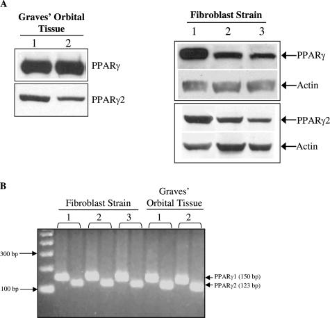
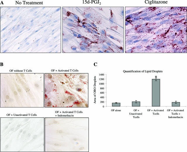
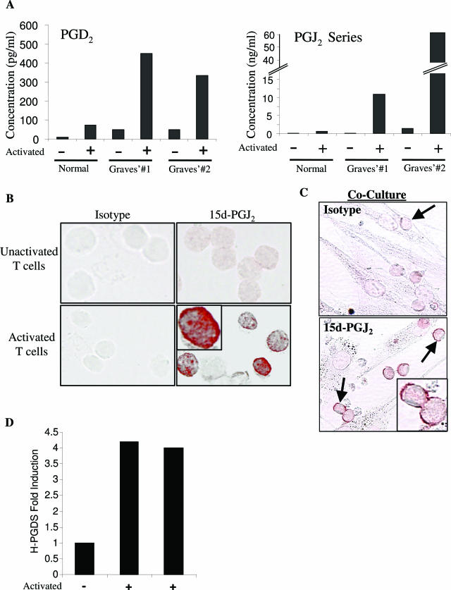
The differentiation of preadipocyte fibroblasts to adipocytes is a crucial process to many disease states including obesity, cardiovascular, and autoimmune diseases. In Graves' disease, the orbit of the eye can become severely inflamed and infiltrated with T lymphocytes as part of the autoimmune process. The orbital fibroblasts convert to fat-like cells causing the eye to protrude, which is disfiguring and can lead to blindness. Recently, the transcription factor peroxisome proliferator activated receptor (PPAR)-gamma and its natural (15d-PGJ2) and synthetic (thiazolidinedione-type) PPAR-gamma agonists have been shown to be crucial to the in vitro differentiation of preadipocyte fibroblasts to adipocytes. We show herein several novel findings. First, that activated T lymphocytes from Graves' patients drive the differentiation of PPAR-gamma-expressing orbital fibroblasts to adipocytes. Second, this adipogenic differentiation is blocked by nonselective small molecule cyclooxygenase (Cox)-1/Cox-2 inhibitors and by Cox-2 selective inhibitors. Third, activated, but not naïve, human T cells highly express Cox-2 and synthesize prostaglandin D2 and related prostaglandins that are PPAR-gamma ligands. These provocative new findings provide evidence for how activated T lymphocytes, through production of PPAR-gamma ligands, profoundly influence human fibroblast differentiation to adipocytes. They also suggest the possibility that, in addition to the orbit, T lymphocytes influence the deposition of fat in other tissues.


Figures

FIGURE 1
FIGURE 1
Human Graves’ orbital fibroblasts express PPAR-γ protein and mRNA. A: A Western blot for total PPAR-γ (both γ1 and γ2 isoforms) and for PPAR-γ2 (using a monoclonal antibody specific for the unique N terminus of PPAR-γ2) was performed on orbital tissue from decompression surgery of two patients and orbital fibroblasts from three different Graves’ patients. Five μg of protein were loaded for each lane and the membranes were reprobed for total actin as a loading control (for fibroblasts only). B: RT-PCR for PPAR-γ1 and PPAR-γ2 was performed on 0.5 μg of RNA from Graves’ orbital decompression tissue (two patients) and orbital fibroblasts (three patients).
FIGURE 2
FIGURE 2
Activated human T lymphocytes drive human Graves’ orbital fibroblasts to adipocytes. A and B: Oil Red O staining was performed on 8-day cultures of orbital fibroblasts exposed to the PPAR-γ agonists 15d-PGJ2 or ciglitazone (A) or co-cultured with activated (rIL-2 and PHA) or unactivated peripheral blood T cells from Graves’ patients (B). B: The Cox inhibitor indomethacin blocked T-cell-induced adipocyte differentiation of orbital fibroblasts. C: The amount of lipid droplet formation for the T-cell orbital fibroblast co-culture was quantified using Image-Pro Software and was plotted as area of lipid droplet formation. *P < 0.007 for orbital fibroblast (OF) plus T cells versus OF alone.
FIGURE 3
FIGURE 3
T-lymphocyte-induced fibroblast differentiation is a Cox-dependent process. Orbital fibroblasts were co-cultured with either activated or unactivated T cells in the presence or absence of vehicle, or the Cox-1/Cox-2 inhibitor indomethacin, or with the Cox-2-specific inhibitor NS-398. After 8 days of co-culture, the triglyceride content of the orbital fibroblasts was measured in a colorimetric triglyceride assay. *P < 0.01 for orbital fibroblasts cultured with activated T cells versus co-cultures treated with Cox inhibitors.
FIGURE 4
FIGURE 4
Activated human T lymphocytes from Graves’ patient peripheral blood strongly express Cox-2. A: A Western blot for Cox-1, Cox-2, and total actin was performed on activated and unactivated T cells from two Graves’ patients. Densitometry was performed to calculate the relative intensities of Cox-1 and Cox-2 expression. B: A Western blot for Cox-1 and Cox-2 was performed on equal amounts of protein from Graves’ orbital tissue (two patients) obtained from decompression surgery.
FIGURE 5
FIGURE 5
Activated, but not unactivated T lymphocytes from Graves’ patients are positive for Cox-2 in co-culture with human orbital fibroblasts. Immunohistochemistry was performed for Cox-2 on 8-day co-cultures of activated (top) or unactivated (bottom) Graves’ T cells with orbital fibroblasts. The activated T cells (arrows) are highly positive for Cox-2, whereas the fibroblasts are either weakly positive or negative for Cox-2.
FIGURE 6
FIGURE 6
Fibroblast-T cell contact is not needed for adipogenesis. Fibroblasts and T cells were either co-cultured in a transwell system in which the fibroblasts and T cells were separated by a 0.4-μm membrane or cultured without separation. 15d-PGJ2 (5 μmol/L) was added as a positive control. After 8 days of culture, the orbital fibroblasts in the transwell system were still able to differentiate to adipocytes as determined by Oil Red O staining.
FIGURE 7
FIGURE 7
Activated human T lymphocytes produce PGD2 and its PGJ2 metabolites, including the PPAR-γ ligand 15d-PGJ2. A: Normal or Graves’ T cells were either activated or not activated for 8 days. PGD2 (left) and the PGJ2 series of PGD2 metabolites, including 15d-PGJ2 (right) were measured in cell culture supernatants by enzyme immunoassay. B: Unactivated or activated Graves’ T cells were stained with a monoclonal anti-15d-PGJ2 antibody. Only activated T cells stained positive for 15d-PGJ2. The isotype-stained cells are negative. C: Graves’ T cells co-cultured with orbital fibroblasts for 3 days stain positive for 15d-PGJ2. The orbital fibroblasts are negative for 15d-PGJ2. The arrows indicate T cells. These experiments were repeated three times with T cells from different Graves’ patients. D: Quantitative RT-PCR for H-PGDS was performed on RNA from unactivated Graves’ T cells (one patient) and activated T cells from two Graves’ patients. Cycle threshold values were normalized to 18S ribosomal RNA, and fold induction over unactivated T cells was calculated. Original magnifications: ×600 (B, C); ×1000 (insets).
FIGURE 8
FIGURE 8
Prostaglandin and T-lymphocyte-induced fibroblast differentiation to adipocytes is a PPAR-γ-dependent process. A: Graves’ orbital fibroblasts were exposed to PGD2 (5 μmol/L), 15d-PGD2 (5 μmol/L), and 15d-PGJ2 (5 μmol/L) with or without the irreversible PPAR-γ antagonist GW9662 (1 μmol/L) for 8 days at which time an assay to measure triglyceride content was performed. The data are the mean of triplicates ± the SD. *P < 0.001 compared with PGs alone. B: Graves’ orbital fibroblasts were co-transfected with the PPRE-LUC reporter construct and a GFP control plasmid, and then co-cultured with Graves’ T cells. 15d-PGJ2 was used as a positive control PPAR-γ ligand. A luciferase assay was performed after 2 days of co-culture. The data are the mean of triplicate samples (±SD), and the luciferase results were plotted as fold increase over the untreated control. *P < 0.05 versus untreated OF cells. C: The irreversible PPAR-γ antagonist GW9662 inhibits the ability of activated T cells to induce adipogenesis in orbital fibroblasts. Fibroblasts were co-cultured with 1 μmol/L GW9662 and activated T cells for 8 days. The results of a triglyceride assay are shown. The data are the mean of triplicates ± the SD. *P < 0.001 versus orbital fibroblasts (OF) plus T cells. There was no statistical difference between OF alone and OF plus T cells plus GW9662.

Similar articles

Cited by

References

    1. Smith RS, Smith TJ, Blieden TM, Phipps RP. Fibroblasts as sentinel cells. Synthesis of chemokines and regulation of inflammation. Am J Pathol. 1997;151:317–322. - PMC - PubMed
    1. Gabbiani G. The myofibroblast: a key cell for wound healing and fibrocontractive diseases. Prog Clin Biol Res. 1981;54:183–194. - PubMed
    1. Phan SH. The myofibroblast in pulmonary fibrosis. Chest. 2002;122:286S–289S. - PubMed
    1. Brody JS, Kaplan NB. Proliferation of alveolar interstitial cells during postnatal lung growth. Evidence for two distinct populations of pulmonary fibroblasts. Am Rev Respir Dis. 1983;127:763–770. - PubMed
    1. Koumas L, Smith TJ, Feldon S, Blumberg N, Phipps RP. Thy-1 expression in human fibroblast subsets defines myofibroblastic or lipofibroblastic phenotypes. Am J Pathol. 2003;163:1291–1300. - PMC - PubMed

Publication types

MeSH terms