. 2006 Oct;169(4):1312-27.
doi: 10.2353/ajpath.2006.051333.

Impact of the loss of Hoxa5 function on lung alveogenesis

Affiliations

Impact of the loss of Hoxa5 function on lung alveogenesis

Isabel Mandeville et al. Am J Pathol. 2006 Oct.

Abstract

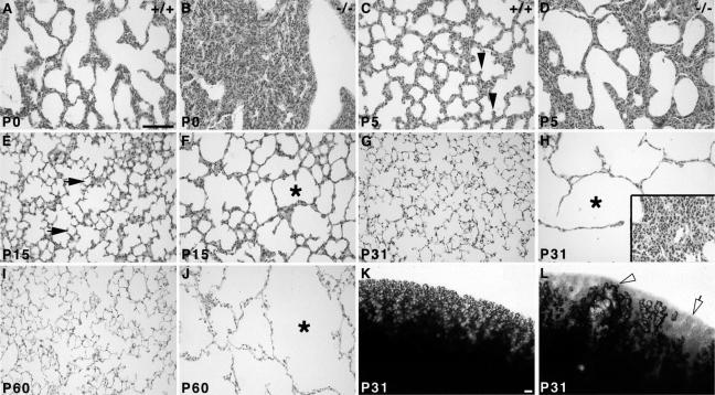
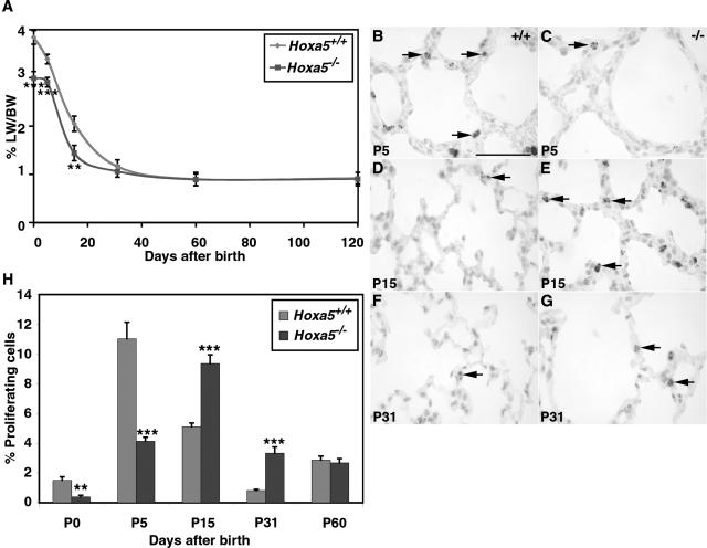
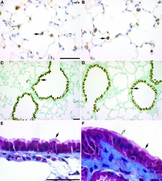
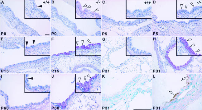
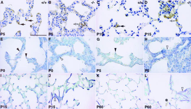
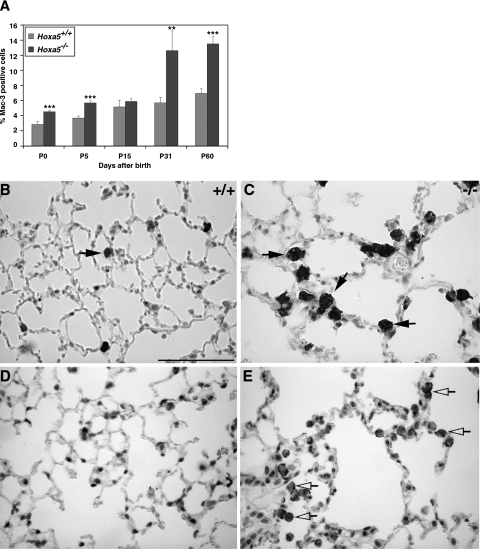
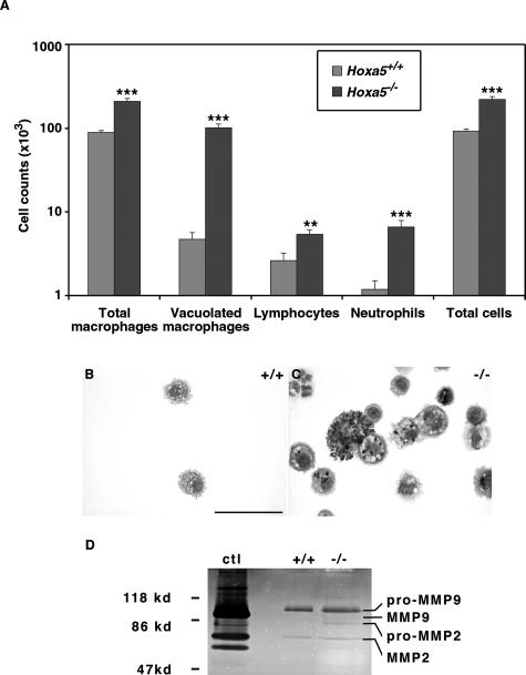
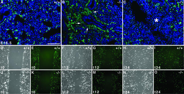
The involvement of genes controlling embryonic processes in the etiology of diseases often escapes attention because of the focus given to their inherent developmental role. Hoxa5 belongs to the Hox gene family encoding transcription factors known for their role in skeletal patterning. Hoxa5 is required for embryonic respiratory tract morphogenesis. We now show that the loss of Hoxa5 function has severe repercussions on postnatal lung development. Hoxa5-/- lungs present an emphysema-like morphology because of impaired alveogenesis. Chronic inflammation characteristics, including goblet cell hyperplasia, mucus hypersecretion, and recruitment of inflammatory cells, were also observed. Altered cell specification during lung morphogenesis triggered goblet cell anomalies. In addition, the defective motility of alveolar myofibroblast precursors in the embryonic lung led to the mispositioning of the alveolar myofibroblasts and to abnormal elastin deposition postnatally. Both goblet cell hyperplasia and elastic fiber abnormalities contributed to the chronic physiopathological features of Hoxa5-/- lungs. They constituted an attractive stimulus to recruit activated macrophages that in turn generated a positive feedback loop that perpetuated macrophage accumulation in the lung. The present work corroborates the notion that altered Hox gene expression may predispose to lung pathologies.


Figures

FIGURE 1
FIGURE 1
Comparative histology (A–J) and macroscopic analysis (K, L) of postnatal wild-type and Hoxa5−/− lungs. At P0, mutant lungs were collapsed in contrast to wild-type specimens (A, B). Alveogenesis initiated normally at P5 in control samples (C, arrowheads), whereas in Hoxa5−/− lungs, the parenchyma was thickened and few septa were noticed (D). At P15, alveoli were readily observed in controls (E, arrows), but Hoxa5−/− lungs rather displayed enlarged airspaces (F, asterisk). At P31 (G, H, K, L) and P60 (I, J), Hoxa5−/− lungs presented an emphysema-like phenotype (H, J, L) with areas depleted of alveoli (open arrow and asterisk) or large tubular structures (open arrowhead), whereas other regions were atelectatic (inset in H) when compared with the normal lung architecture (G, I, K). Scale bars = 100 μm.
FIGURE 2
FIGURE 2
Lung weight/body weight ratio (A) and proliferation rate (B–H) of wild-type and Hoxa5−/− lungs. A: The lung weight/body weight (LW/BW) ratio of Hoxa5−/− mutants was statistically lower than that of controls at P0, P5, and P15. However, a compensatory effect was observed from P31 onwards. Detection of proliferating cells with an anti-phosphorylated histone H3 antibody (B–G, arrows) showed that proliferation was lower in Hoxa5−/− specimens compared with wild-type lungs at P0 (H) and P5 (B, C, H). At P15 (D, E, H) and P31 (F–H), more proliferating cells were detected in mutant lungs than in controls. H: No difference was observed at P60 between both groups. Statistically significant differences are denoted by asterisks (**P < 0.0075, ***P < 0.0006). Scale bar = 50 μm.
FIGURE 3
FIGURE 3
Correct specification of type II pneumocytes (A, B), Clara cells (C, D), and ciliated cells (E, F) in Hoxa5−/− lungs. No major difference was detectable between wild-type and Hoxa5−/− lungs regarding type II pneumocytes and Clara cells as shown by immunostaining with SP-B (A, B) and CC-10 (C, D) antibodies specific for these cell types, respectively (black arrows, shown for P60). Masson’s trichrome revealed that ciliated cells (black arrows) were similarly distributed in mutant (E) and wild-type lungs (F). However, mucus-producing goblet cells were more often visualized in mutant samples (F, open arrow). Scale bars = 50 μm.
FIGURE 4
FIGURE 4
Goblet cell abnormal distribution in Hoxa5−/− lungs. Goblet cells were revealed by periodic acid/Schiff staining in postnatal lungs (A–J). At all ages tested, only a few positive cells were detected in wild-type samples (A, C, E, G, I; arrowheads), mostly scattered along the epithelium of the trachea (A, C) and the primary bronchi (E, I). No goblet cell was detected distally to the primary bronchi in controls (G). In Hoxa5−/− lungs, an increase in goblet cell number was systematically observed at every stage (B, D, F, H, J; open arrowheads), most obviously from P15 onwards (F, H, J). Furthermore, positive cells were detected in more distal structures such as the secondary bronchi (H) and bronchioles (not shown). Mucin MUC5AC, a marker of mucus hypersecretion, was detected by immunostaining (K, L). Although control samples remained negative (K), positive cells stained at their apical side were observed along the bronchi of Hoxa5−/− specimens from P15 onwards (shown for P31; L, open arrows). Scale bars = 50 μm.
FIGURE 5
FIGURE 5
Perturbed alveolar myofibroblasts localization (A–D) and elastin deposition (E–L) in Hoxa5−/− lungs. Alveolar myofibroblasts were located at the tip of the septa of control specimens at P5 and P15, as revealed by immunostaining with an α-SMA antibody (A, C; arrows). In Hoxa5−/− lungs, they were trapped within the parenchyma (B, D; open arrow). Weigert staining allowed visualization of elastic fibers (E–L). In P0 and P5 control specimens, elastic fibers were localized along the respiratory saccules and at the tip of the growing septa (E, G; arrowheads). In Hoxa5−/− lungs, elastin appeared tangled in the parenchyma (F, H; open arrowhead). At P15 onwards, Hoxa5−/− lungs displayed disorganized fibers (J, L; open arrowhead) compared with wild-type lungs (I, K). Moreover, inflammatory cells were detected in P60 mutant lungs (L, asterisk). Scale bar = 50 μm.
FIGURE 6
FIGURE 6
Increased percentage of alveolar macrophages in Hoxa5−/− lungs. A Mac-3-specific antibody was used to detect macrophages in the lung (A–C, arrows) showing that the proportion of macrophages was statistically higher in Hoxa5−/− lungs than in controls. Asterisks denote statistical differences (**P < 0.01, ***P < 0.005). C: Moreover, at P31 (not shown) and P60, accumulation of large vacuolated macrophages was observed in Hoxa5−/− lungs. MMP-12 immunostaining revealed that the majority of macrophages in mutant samples produced this elastase (shown for P60; D and E, open arrows). Scale bar = 50 μm.
FIGURE 7
FIGURE 7
Increased number of inflammatory cells and MMP activity in Hoxa5−/− BALF. A: Differential cell counts showed an increase of macrophages, lymphocytes, and neutrophils in Hoxa5−/− specimens when compared with controls. Statistically significant differences are denoted by asterisks (**P < 0.002, ***P < 0.0001). Diff-Quik staining also revealed that Hoxa5−/− BALF contained large, vacuolated macrophages with dark inclusions not seen in control samples (B, C). D: MMP activities in wild-type (+/+) and Hoxa5−/− (−/−) alveolar macrophage-conditioned media in gelatin gel zymogram. Data are presented as the reverse image of the original zymogram. Conditioned media from the HT1080 cell line served as a positive control (ctl). Slightly elevated MMP2 and MMP9 activities were observed in Hoxa5−/− alveolar macrophage-conditioned medium. Scale bar = 50 μm.
FIGURE 8
FIGURE 8
Defective motility of alveolar myofibroblast progenitors. A–C: Visualization of the GFP-positive alveolar myofibroblasts progenitors in E16.5 wild-type lungs showed that progenitors were dispersed in the lung parenchyma (A). In contrast, most Pdgfαr/GFP-positive cells remained clustered around the epithelium in Hoxa5−/− lungs, indicating a block in distal spreading of the alveolar myofibroblast progenitors (B, arrows). Progenitors also failed to invade some lung regions (C, asterisk). Sections were counterstained with DAPI in blue. D–O: Wounding assays of primary mesenchymal cells isolated from lungs of E15.5 Hoxa5+/+;PdgfαrGFP/+ (D–I) and Hoxa5−/−;PdgfαrGFP/+ (J–O) embryos after 0 (t0; D, E, J, K), 12 (t12; F, G, L, M), and 24 hours (t24; H, I, N, O). Wild-type cells invaded the wounded area within 12 hours of the assay (F, G), filling the gap by t24 (H, I). In contrast Hoxa5−/−;PdgfαrGFP/+ cells showed reduced migration at both time points (L–O). Scale bars = 50 μm.
FIGURE 9
FIGURE 9
Hoxa5 protein detection in primary embryonic lung myofibroblasts. A: Mesenchymal cells isolated from E15.5 lungs adopted a myofibroblast phenotype as shown by the co-expression of Pdgf-α receptor/GFP and α-SMA. B: The actin staining observed with phalloidin was identical to that of α-SMA in GFP-positive cells. C and D: The Hoxa5 protein was readily detected in the nucleus of wild-type myofibroblasts. D, E, and G: Furthermore, GFP and Hoxa5 protein fluorescence extensively overlapped in this cell population. H–K: The Hoxa5 protein was not detected in Hoxa5−/− cell cultures. Nuclei were counterstained with DAPI (A, C, F, G, J, K). Scale bar = 100 μm.

Similar articles

Cited by

References

    1. Perl A-K, Whitsett JA. Molecular mechanisms controlling lung morphogenesis. Clin Genet. 1999;56:14–27. - PubMed
    1. Cardoso WV. Lung morphogenesis revisited: old facts, current ideas. Dev Dyn. 2000;219:121–130. - PubMed
    1. Warburton D, Schwarz M, Tefft D, Flores-Delgado G, Anderson KD, Cardoso WV. The molecular basis of lung morphogenesis. Mech Dev. 2000;15:55–81. - PubMed
    1. Rogers DF. Mucus hypersecretion in chronic obstructive pulmonary disease. Chronic obstructive pulmonary disease: pathogenesis to treatment. Novartis Symposium. 2001;234:65–83. - PubMed
    1. Barnes PJ. Small airways in COPD. N Engl J Med. 2004;350:2635–2637. - PubMed

Publication types

MeSH terms