. 2006 Oct;169(4):1458-72.
doi: 10.2353/ajpath.2006.051358.

Recruitment of a prostaglandin E receptor subtype, EP3-expressing bone marrow cells is crucial in wound-induced angiogenesis

Affiliations

Recruitment of a prostaglandin E receptor subtype, EP3-expressing bone marrow cells is crucial in wound-induced angiogenesis

Emi Kamoshita et al. Am J Pathol. 2006 Oct.

Abstract

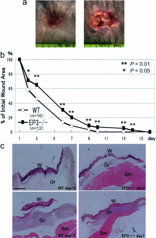
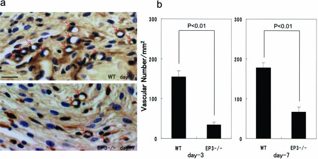
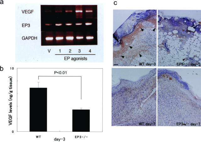
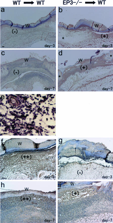
E-type prostaglandins have been reported to be proangiogenic in vivo. Thus, we examined prostaglandin receptor signaling relevant to wound-induced angiogenesis. Full-thickness skin wounds were created on the backs of mice, and angiogenesis in wound granulation tissues was estimated. Wound closure and re-epithelization in EP3 receptor knockout mice (EP3-/-) were significantly delayed compared with their wild-type (WT) mice, whereas those in EP1-/-, EP2-/-, and EP4-/- were not delayed. Wound-induced angiogenesis estimated with CD31 immunohistochemistry in EP3-/- mice was significantly inhibited compared with that in WT mice. Immunoreactive vascular endothelial growth factor (VEGF) in wound granulation tissues in EP3-/- mice was markedly less than that in WT mice. Wound closure in WT mice was delayed significantly by VEGF neutralizing antibody compared with control IgG. Wound-induced angiogenesis and wound closure were significantly suppressed in EP3-/- bone marrow transplantation mice compared with those in WT bone marrow transplantation mice. These were accompanied with the reductions in accumulation of VEGF-expressing cells in wound granulation tissues and in mobilization of VEGF receptor 1-expressing leukocytes in peripheral circulation. These results indicate that the recruitment of EP3-expressing cells to wound granulation tissues is critical for surgical wound healing and angiogenesis via up-regulation of VEGF.


Figures

FIGURE 1
FIGURE 1
Delayed wound healing in EP3 receptor knockout mice. Surgical wounds were made on the backs of EP3 receptor knockout mice (EP3−/−) and their WT counterparts, and wound closure was determined as described in Materials and Methods. a: Typical appearance of wounds in EP3−/− and WT at day 7. The original diameter of the wounds was 8 mm. One division on the scale below the wound represents 1 mm. b: Time course of wound closure in EP3−/− and WT. Data are means ± SEM for the indicated number of mice. *P < 0.05, and **P < 0.01 versus WT mice (analysis of variance). c: H&E staining was used for wound tissues including granulation tissues from EP3−/− and WT. Tissues were fixed at days 3 and 7. W, wound; Gr, granulation tissue; Sm, skeletal muscle. Scale bar = 1 mm.
FIGURE 2
FIGURE 2
Angiogenesis in wound granulation tissues in EP3 receptor knockout mice. Surgical wounds were made on the backs of EP3 receptor knockout mice (EP3−/−) and of their WT counterparts, and angiogenesis in the wound granulation tissues was determined as described in Materials and Methods. a: Typical results of CD31 immunostaining in the wound granulation tissues in EP3−/− and WT at day 3. CD-31-positive microvessels (brown stains indicated by arrowheads) were rich in the granulation tissues in WT but were apparently poor in EP3−/−. Arrowheads in a indicate microvessel-like constructions. b: Changes in the microvessel density in the granulation tissues at days 3 and 7. After CD-31 immunohistochemistry was photographed, the microvessel density per observation field (100 μm × 100 μm) of the wound granulation tissues was counted. The microvessel density was finally expressed per mm2, and results from 10 100 μm × 100 μm fields were averaged. Data are means ± SEM for six mice. Analysis of variance was used to test the significance of difference. Scale bar = 50 μm.
FIGURE 3
FIGURE 3
Expression of VEGF in sponge granulation tissues in WT mice stimulated with selective EP agonists and in wound granulation tissues in EP3 receptor knockout mice. a: VEGF expressions in sponge granulation tissues in WT mice stimulated with selective EP agonists for 2 weeks. EP receptor subtype-selective agonists, ONO-DI-004, ONO-AEI-257, ONO-AE-248, and ONO-AEI-329, which were specific to EP1, EP2, EP3, and EP4, respectively, were topically injected (15 nmol/sponge, twice a day) into the sponges implanted in the subcutaneous tissues of WT mice. RT-PCR was performed as described in the text. b: Immunoreactive VEGF levels, determined by ELISA, in the wound granulation tissues in EP3−/− mice and WT mice at day 3. VEGF protein was not detected in normal skin tissue in WT mice (data not shown). VEGF expression was detected in wound tissue in WT mice (left) but was reduced in EP3−/− mice (right). To test the significance of difference, t-test was used. c: Typical results of VEGF immunostaining in the wound granulation tissues in EP3−/− and WT at days 3 and 7. VEGF-positive cells were markedly accumulated in the wound granulation tissues in WT mice at day 3 (brown stains indicated by arrowheads). Scale bar = 100 μm.
FIGURE 4
FIGURE 4
Time course of wound healing and angiogenesis in mice transplanted with BM cells from EP3 receptor knockout mice. A lethal dose of radiation was given to WT mice, and BM cells either from WT mice or from EP3−/− mice were injected into the tail vein under light ether anesthesia (WT→WT or EP3−/−→WT). Surgical wounds were made on the backs of the mice, and wound closure was determined as described in Materials and Methods. a: Time course of wound closure in WT mice transplanted with BM cells either from EP3−/− or from WT mice. Data are means ± SEM for the indicated number of mice. *P < 0.05 versus WT→WT mice (analysis of variance). b: The changes in the microvessel density in the wound granulation tissues in WT mice transplanted with BM cells either from EP3−/− or from WT mice. After CD31 immunohistochemistry was photographed, the number of vessels in the observed area was counted and expressed per mm2 observed. Data are means ± SEM for the indicated number of mice. P < 0.05 versus WT→WT (analysis of variance). c: Expressions of EP receptor subtypes in BM cells isolated from EP3−/− mice and WT mice. RT-PCR was performed as described in the text.
FIGURE 5
FIGURE 5
Typical results of immunostainings of β-galactosidase (a–d) and VEGF (f–i) and of EP3 in situ hybridization (e) in the wound granulation tissues in WT mice transplanted with BM cells either from EP3−/− mice (EP3−/−→WT) or from WT mice (WT→WT) at day 3 (a, b, eg) and at day 7 (c, d, h, and i). β-Galactosidase-positive cells accumulated in the wound granulation tissues in WT mice transplanted with BM cells from EP3−/− mice (b and d, ++). WT mice receiving WT BM cells did not show β-galactosidase-positive stains in the wound tissues (a and c). The uninjured lesions around the wounds introduced in WT mice transplanted with BM cells from EP3−/− mice did not exhibit β-galactosidase (b and d, asterisks). EP3 mRNA was detected in granulation tissues just beneath the wound in WT mice receiving WT BM cells (e, high magnitude). VEGF immunoreactivity was mainly seen in granulation tissues in WT mice receiving WT BM cells (f and h), whereas no marked accumulation of VEGF-positive cells was seen in WT mice implanted with EP3−/− cells (g and i). W indicates surgical wound in recovery stage. Scale bars = 100 μm (a, f); 25 μm (e).

References

    1. Levi S, Goodlad RA, Lee CY, Stamp G, Walport MJ, Wright NA, Hodgson HJ. Inhibitory effect of non-steroidal anti-inflammatory drugs on mucosal cell proliferation associated with gastric ulcer healing. Lancet. 1990;336:840–843. - PubMed
    1. Wang JY, Yamasaki S, Takeuchi K, Okabe S. Delayed healing of acetic acid-induced gastric ulcers in rats by indomethacin. Gastroenterology. 1989;96:393–402. - PubMed
    1. Tarnawski A, Stachura J, Douglass TG, Krause WJ, Gergely H, Sarfeh IJ. Indomethacin impairs quality of experimental gastric ulcer healing: a quality histologic and ultrastructural analysis. Garner A, O’Brien PE, editors. Chichester: J Wiley & Sons,; Mechanisms of Injury, Protection and Repair of the Upper Gastrointestinal Tract. 1991:pp 521–531.
    1. Schmassmann A, Tarnawski A, Peskar BM, Varga L, Flogerzi B, Halter F. Influence of acid and angiogenesis on kinetics of gastric ulcer healing in rats: interaction with indomethacin. Am J Physiol. 1995;268:G276–G285. - PubMed
    1. Tarnawski A. Cellular mechanisms of gastric ulcer healing. Domschke W, Konturek SJ, editors. New York: Springer-Verlag,; The Stomach. 1993:pp 177–192.

Publication types

MeSH terms

Substances

LinkOut - more resources