. 2006 Oct;169(4):1484-95.
doi: 10.2353/ajpath.2006.060116.

Influence of endothelial cells on vascular smooth muscle cells phenotype after irradiation: implication in radiation-induced vascular damages

Affiliations

Influence of endothelial cells on vascular smooth muscle cells phenotype after irradiation: implication in radiation-induced vascular damages

Fabien Milliat et al. Am J Pathol. 2006 Oct.

Abstract

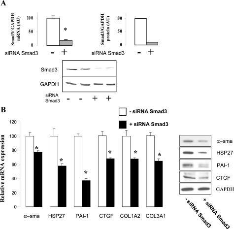
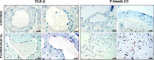
Damage to vessels is one of the most common effects of therapeutic irradiation on normal tissues. We undertook a study in patients treated with preoperative radiotherapy and demonstrated in vivo the importance of proliferation, migration, and fibrogenic phenotype of vascular smooth muscle cells (VSMCs) in radiation-induced vascular damage. These lesions may result from imbalance in the cross talk between endothelial cells (ECs) and VSMCs. Using co-culture models, we examined whether ECs influence proliferation, migration, and fibrogenic phenotype of VSMCs. In the presence of irradiated ECs, proliferation and migration of VSMCs were increased. Moreover, expressions of alpha-smooth muscle actin, connective tissue growth factor, plasminogen activator inhibitor type 1, heat shock protein 27, and collagen type III, alpha 1 were up-regulated in VSMCs exposed to irradiated ECs. Secretion of transforming growth factor (TGF)-beta1 was increased after irradiation of ECs, and irradiated ECs activated the Smad pathway in VSMCs by inducing Smad3/4 nuclear translocation and Smad-dependent promoter activation. Using small interferring RNA targeting Smad3 and a TGFbeta-RII neutralizing antibody, we demonstrate that a TGF-beta1/TGF-beta-RII/Smad3 pathway is involved in the fibrogenic phenotype of VSMCs induced by irradiated ECs. In conclusion, we show the importance of proliferation, migration, and fibrogenic phenotype of VSMCs in patients. Moreover, we demonstrate in vitro that ECs influence these fundamental mechanisms involved in radiation-induced vascular damages.


Figures

FIGURE 1
FIGURE 1
Characterization of radiation-induced vascular damages. A: Radiation injury score and vessels morphometric measurements (the ratio between luminal surface and outer surface) were performed in tissues from 38 patients treated by radiotherapy for rectal adenocarcinoma. Values of radiation injury score for every point constitute the sum (the retrieved sum to 100) of the score of every parameter observed in each compartment for one slide. B: Representative microscopic images from control (A, E, I, M, Q, U) and irradiated (B–D, F–H, J–L, N–P, R–T, V–X) submucosal vessels: H&E coloration (A–D), Masson’s trichrome (E–H), elastin coloration (I–J); and immunolabeling of α-SMA (K–K′), calponin (L–L′), collagen I (M–P), collagen III (Q–T), and PCNA (U–X, arrows indicate some PCNA-positive cells) are shown. C: Representative microscopic images from control lung, uterus, and skin (A, B), and irradiated (C–D) tissue are shown (Meyer’s hemalum coloration).
FIGURE 2
FIGURE 2
Irradiated ECs influence cell cycle progression and VSMC proliferation. In all experiments, 50% confluent VSMCs were serum-starved for 24 hours before co-culture and irradiation. VSMCs were changed with complete culture medium, and Transwell-containing confluent ECs were incubated with VSMCs and then irradiated. A: Proliferation of VSMCs was determined by cell counting. Data are the mean ± SEM of three experiments realized in triplicate or quadruplicate. * or # or §P < 0.05 versus VSMCs; values with different symbols are statistically different. B: Representative cultures obtained 2 days after irradiation are shown. C: Cell cycle distribution was determined by propidium iodide staining. Data are the mean ± SEM, and for each time, values with different footnote letters are statistically different from each other (P < 0.05).
FIGURE 3
FIGURE 3
Irradiated ECs induce VSMC migration. Migration of VSMCs was determined by the scratch injury model. ECs and VSMCs were cultured separately at confluence. Just before co-culture and irradiation, confluent VSMCs were scratched (80 μm) by a regular pipette tip (three wounds per well) and rinsed, and the culture medium remained unchanged during wound healing. Transwell-containing confluent ECs were incubated to confluent VSMCs just before irradiation (10 Gy), when the two cell types are co-irradiated or just after irradiation when ECs or VSMCs are irradiated. A: Representative images of VSMCs at days 1 and 4 from two separate experiments realized in duplicate or triplicate. B: Migration index was determined 1, 2, and 4 days after irradiation as described in Materials and Methods. Migration index is the mean ± SEM of two experiments realized in triplicate. *P < 0.05 versus VSMCs alone; #P < 0.05 versus VSMCs, 10 Gy + ECs.
FIGURE 4
FIGURE 4
Irradiated ECs induce a VSMC fibrogenic phenotype. ECs and VSMCs were cultured separately at confluence and settled together at the moment of co-culture and irradiation (2 or 10 Gy). Fibrogenic phenotype of VSMCs was investigated by real-time PCR (24 hours after irradiation) and Western blot (48 hours after irradiation). A: Co-culture of irradiated ECs in the presence of irradiated VSMCs at the same dose. B: VSMCs irradiated alone to investigate direct radiation effects. C: Co-culture of irradiated ECs in the presence of nonirradiated VSMCs. Representative Western blots. Data are the mean ± SEM of two to four independent experiments realized in duplicate or triplicate. *P < 0.05 versus control.
FIGURE 5
FIGURE 5
Irradiation increases TGF-β1 secretion in ECs. Total and active TGF-β1 contents were determined by ELISA assay in EC and VSMC supernatants 24 hours after 2 or 10 Gy irradiation. Data are the mean ± SEM of three experiments realized in triplicate. *P < 0.05 versus control.
FIGURE 6
FIGURE 6
Irradiated ECs activate Smad pathway in VSMCs. A: ECs induce the nuclear translocation of Smad3 and Smad4 in VSMCs after irradiation. VSMCs and ECs were cultured separately at confluence. VSMCs were incubated in serum-free medium for 24 hours before experiment. Just before co-culture and irradiation (10Gy), ECs were changed in complete medium and VSMCs with serum-free medium. Twenty-four hours after irradiation, Smad3 and Smad4 nuclear translocation in VSMCs was followed by immunofluorescence and examined by confocal microscopy. Representative immunostainings of three independent observations are shown, as well as staining of VSMCs treated with 10 ng/ml TGF-β1 for 1 hour. B: ECs induce a Smad-dependent transcription in VSMCs. VSMCs (50% confluent) were transiently cotransfected in complete medium with (CAGA)9-Lux reporter (1 μg) and pRL-TK (0.2 μg) plasmids using FuGENE 6 (Roche Diagnostics) as transfection reagent (3 μl/1.2 μg of DNA). Twenty-four hours after transfection, VSMCs were changed with serum-free medium then incubated with confluent ECs. Relative luciferase activity (ratio Firefly/Renilla) was measured 24 hours after co-culture and irradiation. Transfection efficiency (about 40%) was estimated using pEGFP-N1 vector (Clontech, Mountain View, CA). VSMCs treated by 3 ng/ml TGF-β1 for 24 hours are shown. Data are the mean ± SEM (n = 6) *P < 0.05 versus VSMCs alone.
FIGURE 7
FIGURE 7
Smad3 is involved in fibrogenic phenotype of VSMCs induced by ECs after irradiation of both cell types. VSMCs were transfected with cytofectin (1 μg/ml) and 100 nmol/L siRNAs targeting Smad3. A: The silencing efficiency was determined by real-time PCR and Western blot. B: VSMCs and ECs were cultured separately at confluence. VSMCs were transfected 24 hours before co-culture and irradiation of both cell types (10 Gy). Just before irradiation, ECs were changed in complete medium and VSMCs with complete medium ± siRNA Smad3 transfection solution. Fibrogenic phenotype of VSMCs was investigated by real-time PCR (24 hours after irradiation) and Western blot (48 hours after irradiation). Data are the mean ± SEM of two experiments realized in triplicate. *P < 0.05 versus 10 Gy irradiated cells without siRNA Smad3. Representative Western blots are shown with irradiation of both cell types at 10 Gy ± siRNA Smad3.
FIGURE 8
FIGURE 8
TGFβ-RII is involved in fibrogenic phenotype of VSMCs induced by ECs after irradiation of both cell types. VSMCs and ECs were cultured separately at confluence. A: VSMCs were serum-starved during 24 hours and preincubated 2 hours before co-culture and irradiation of both cell types (10 Gy) with a goat anti-human TGFβ-RII neutralizing antibody (10 μg/ml in serum-free medium) or normal goat IgG (10 μg/ml). Just before irradiation, ECs were changed in complete medium and VSMCs with serum-free medium. Twenty-four hours after irradiation, Smad3 and Smad4 nuclear translocations in VSMCs were analyzed by immunofluorescent staining. B: VSMCs were preincubated 2 hours before co-culture and irradiation of both cell types (10 Gy) with a goat anti-human TGFβ-RII or normal goat IgG (10 μg/ml). Just before irradiation, ECs and VSMCs were changed in complete medium. Fibrogenic phenotype of VSMCs was investigated by real-time PCR (24 hours after irradiation) and Western blot (48 hours after irradiation). Data are the mean ± SEM. *P < 0.05 versus 10 Gy-irradiated cells with normal IgG. Representative Western blots are shown with co-irradiation of both cell types at 10 Gy ± goat anti-human TGFβ-RII.
FIGURE 9
FIGURE 9
Overexpression of TGF-β and P-Smad 2/3 in radiation-induced vascular lesions. Immunohistochemical stainings of TGF-β and P-Smad 2/3 were performed in tissues from patients treated by radiotherapy for rectum adenocarcinoma. Representative microscopic images from control and irradiated submucosal vessels are shown.

References

    1. Stone HB, Coleman CN, Anscher MS, McBride WH. Effects of radiation on normal tissue: consequences and mechanisms. Lancet Oncol. 2003;4:529–536. - PubMed
    1. Dorresteijn LD, Kappelle AC, Boogerd W, Klokman WJ, Balm AJ, Keus RB, van Leeuwen FE, Bartelink H. Increased risk of ischemic stroke after radiotherapy on the neck in patients younger than 60 years. J Clin Oncol. 2002;20:282–288. - PubMed
    1. Molla M, Gironella M, Miquel R, Tovar V, Engel P, Biete A, Pique JM, Panes J. Relative roles of ICAM-1 and VCAM-1 in the pathogenesis of experimental radiation-induced intestinal inflammation. Int J Radiat Oncol Biol Phys. 2003;57:264–273. - PubMed
    1. Panes J, Anderson DC, Miyasaka M, Granger DN. Role of leukocyte-endothelial cell adhesion in radiation-induced microvascular dysfunction in rats. Gastroenterology. 1995;108:1761–1769. - PubMed
    1. Molla M, Gironella M, Salas A, Miquel R, Perez-del-Pulgar S, Conill C, Engel P, Biete A, Pique JM, Panes J. Role of P-selectin in radiation-induced intestinal inflammatory damage. Int J Cancer. 2001;96:99–109. - PubMed

Publication types

MeSH terms

LinkOut - more resources