

Mycobacteria lacking the RD1 region do not induce necrosis in the lungs of mice lacking interferon-gamma
- PMID: 17005003
- PMCID: PMC1782352
- DOI: 10.1111/j.1365-2567.2006.02427.x
Mycobacteria lacking the RD1 region do not induce necrosis in the lungs of mice lacking interferon-gamma
Abstract
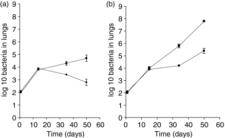
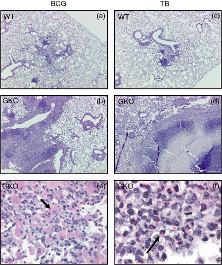
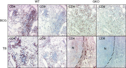
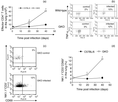
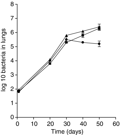
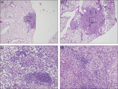
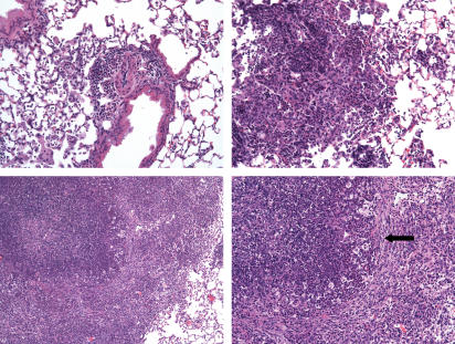
The genetic region of difference 1 (RD1) in Mycobacterium tuberculosis has recently been hypothesized to encode for proteins that are cytotoxic to the host cell in nature. We demonstrate here that while M. tuberculosis grew progressively in the lungs of gene disrupted mice (GKO) unable to produce interferon-gamma (IFN-gamma), similar mice infected instead with M. bovis bacillus Calmette-Guérin (BCG) reproducibly exhibited an obvious slowing of the disease after about 20 days. Closer examination of BCG-infected GKO mice showed a florid granulomatous inflammation in the lungs, whereas similar mice infected with M. tuberculosis exhibited wholesale progressive necrosis. In the BCG-infected GKO mice large numbers of activated effector T cells, some strongly positive for the cytokine tumour necrosis factor, as well as activated natural killer cells accumulated in the lungs. To further test the hypothesis that the differences observed were directly associated with the loss of the RD1 region, it was then shown that a mutant of M. tuberculosis lacking RD1 grew progressively in both normal and GKO mice but failed to induce any degree of necrosis in either animal despite reaching similar levels in the lungs. However, when mice were infected with this mutant, in which the RD1 region had been restored by complementation, wholesale necrosis of the lungs again occurred. These data support the hypothesis that proteins encoded in the RD1 region are a major cause of necrosis and contribute significantly to the pathogenesis of the disease.
Figures
Similar articles
-
Influence of ESAT-6 secretion system 1 (RD1) of Mycobacterium tuberculosis on the interaction between mycobacteria and the host immune system.J Immunol. 2005 Mar 15;174(6):3570-9. doi: 10.4049/jimmunol.174.6.3570. J Immunol. 2005. PMID: 15749894
-
Induction of CD8 T cells against a novel epitope in TB10.4: correlation with mycobacterial virulence and the presence of a functional region of difference-1.J Immunol. 2007 Sep 15;179(6):3973-81. doi: 10.4049/jimmunol.179.6.3973. J Immunol. 2007. PMID: 17785835
-
Mycobacterium tuberculosis H37Rv: Delta RD1 is more virulent than M. bovis bacille Calmette-Guérin in long-term murine infection.J Infect Dis. 2004 Jul 1;190(1):123-6. doi: 10.1086/421472. Epub 2004 Jun 11. J Infect Dis. 2004. PMID: 15195251 Free PMC article.
-
Is interferon-gamma the right marker for bacille Calmette-Guérin-induced immune protection? The missing link in our understanding of tuberculosis immunology.Clin Exp Immunol. 2012 Sep;169(3):213-9. doi: 10.1111/j.1365-2249.2012.04614.x. Clin Exp Immunol. 2012. PMID: 22861360 Free PMC article. Review.
-
Role of M. tuberculosis RD-1 region encoded secretory proteins in protective response and virulence.Tuberculosis (Edinb). 2008 Nov;88(6):510-7. doi: 10.1016/j.tube.2008.05.002. Epub 2008 Jul 21. Tuberculosis (Edinb). 2008. PMID: 18640874 Review.
Cited by
-
Mycobacterium tuberculosis subverts the TLR-2-MyD88 pathway to facilitate its translocation into the cytosol.PLoS One. 2014 Jan 27;9(1):e86886. doi: 10.1371/journal.pone.0086886. eCollection 2014. PLoS One. 2014. PMID: 24475192 Free PMC article.
-
Mouse models of human TB pathology: roles in the analysis of necrosis and the development of host-directed therapies.Semin Immunopathol. 2016 Mar;38(2):221-37. doi: 10.1007/s00281-015-0538-9. Epub 2015 Nov 5. Semin Immunopathol. 2016. PMID: 26542392 Free PMC article. Review.
-
Potential role for ESAT6 in dissemination of M. tuberculosis via human lung epithelial cells.Mol Microbiol. 2010 Jan;75(1):92-106. doi: 10.1111/j.1365-2958.2009.06959.x. Epub 2009 Nov 10. Mol Microbiol. 2010. PMID: 19906174 Free PMC article.
-
Acute immune response to Mycobacterium massiliense in C57BL/6 and BALB/c mice.Infect Immun. 2010 Apr;78(4):1571-81. doi: 10.1128/IAI.00731-09. Epub 2010 Feb 1. Infect Immun. 2010. PMID: 20123718 Free PMC article.
-
Disseminated disease severity as a measure of virulence of Mycobacterium tuberculosis in the guinea pig model.Tuberculosis (Edinb). 2008 Jul;88(4):295-306. doi: 10.1016/j.tube.2007.12.003. Epub 2008 Mar 5. Tuberculosis (Edinb). 2008. PMID: 18321783 Free PMC article.
References
-
- Dye C, Scheele S, Dolin P, Pathania V, Raviglione MC. Consensus statement. Global burden of tuberculosis: estimated incidence, prevalence, and mortality by country. WHO Global Surveillance and Monitoring Project. JAMA. 1999;282:677–86. - PubMed
-
- Raviglione MC, Dye C, Schmidt S, Kochi A. Assessment of worldwide tuberculosis control. WHO Global Surveillance and Monitoring Project. Lancet. 1997;350:624–9. - PubMed
-
- Colditz GA, Brewer TF, Berkey CS, Wilson ME, Burdick E, Fineberg HV, Mosteller F. Efficacy of BCG vaccine in the prevention of tuberculosis. Meta-analysis of the published literature. JAMA. 1994;271:698–702. - PubMed
-
- Kearns AM, Magee JG, Gennery A, Steward M, Graham C, Seiders PR, Freeman R. Rapid identification of Mycobacterium bovis BCG by the detection of the RD1 deletion using a multiplex PCR technique. Int J Tuberc Lung Dis. 1999;3:635–8. - PubMed
Publication types
MeSH terms
Substances
Grants and funding
LinkOut - more resources
Full Text Sources
Medical
Molecular Biology Databases
