Review
. 2006 Oct;209(4):447-68.
doi: 10.1111/j.1469-7580.2006.00635.x.

Soluble and insoluble signals and the induction of bone formation: molecular therapeutics recapitulating development

Affiliations
Review

Soluble and insoluble signals and the induction of bone formation: molecular therapeutics recapitulating development

Ugo Ripamonti et al. J Anat. 2006 Oct.

Abstract

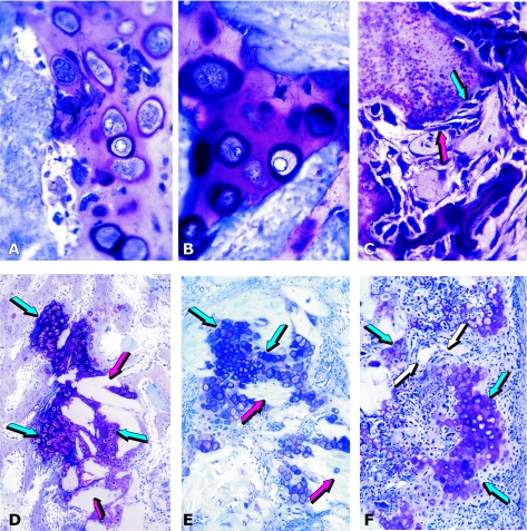
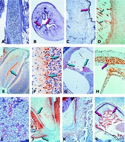
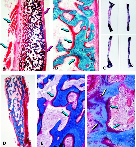
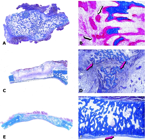
The osteogenic molecular signals of the transforming growth factor-beta (TGF-beta) superfamily, the bone morphogenetic/osteogenic proteins (BMPs/OPs) and uniquely in primates the TGF-beta isoforms per se, pleiotropic members of the TGF-beta supergene family, induce de novo endochondral bone formation as a recapitulation of embryonic development. Naturally derived BMPs/OPs and gamma-irradiated human recombinant osteogenic protein-1 (hOP-1) delivered by allogeneic and xenogeneic insoluble collagenous matrices initiate de novo bone induction in heterotopic and orthotopic sites of the primate Papio ursinus, culminating in complete calvarial regeneration by day 90 and maintaining the regenerated structures by day 365. The induction of bone by hOP-1 in P. ursinus develops as a mosaic structure with distinct spatial and temporal patterns of gene expression of members of the TGF-beta superfamily that singly, synergistically and synchronously initiate and maintain tissue induction and morphogenesis. The temporal and spatial expressions of TGF-beta1 mRNA indicate a specific temporal transcriptional window during which expression of TGF-beta1 is mandatory for successful and optimal osteogenesis. Highly purified naturally derived bovine BMPs/OPs and hOP-1 delivered by human collagenous bone matrices and porous hydroxyapatite, respectively, induce bone formation in mandibular defects of human patients. By using healthy body sites as bioreactors it is possible to recapitulate embryonic developments by inducing selected biomaterials combined with recombinant proteins to transform into custom-made prefabricated bone grafts for human reconstruction. The osteogenic proteins of the TGF-beta superfamily, BMPs/OPs and TGF-betas, the last endowed with the striking prerogative of inducing endochondral bone formation in primates only, are helping to engineer skeletal reconstruction in molecular terms.


Figures

Fig. 1
Fig. 1
Tissue induction and morphogenesis by naturally derived and recombinant human osteogenic proteins delivered by collagenous matrices as carrier in the rodent bioassay. (A,B) Induction of chondrogenesis by 0.1–0.5 µg osteogenin purified to apparent homogeneity after electroendosmotic elution of baboon osteogenic fractions; and (C) osteogenin induces osteoblastic cells secreting bone matrix and capillary invasion with differentiating mesenchymal cells migrating from the vascular compartment (red arrow) to the bone matrix compartment (blue arrow) with secreting osteoblastic cells attached to the matrix. (D–F) Induction of endochondral bone differentiation by 2 µg hOP-1 delivered by insoluble collagenous bone matrix (red arrows) as carrier implanted in the subcutaneous space of the rodent bioassay and harvested on day 11. Islands of cartilage induction as a recapitulation of embryonic development (blue arrows), vascular invasion (white arrows in F), chondrolysis and bone differentiation.
Fig. 2
Fig. 2
Pleiotropism of bone morphogenetic proteins: from bone to central and peripheral nervous system expression and localization. (A) In situ hybridization of OP-1 mRNA in the developing femur of a human embryo. (B) OP-1 mRNA localized in the intestinal epithelium of the human fetus and (C) epidermis (red arrows). (D) Sixteen-day-old pup: BMP-3 immunolocalization in the cerebral cortex delineating neurite axonal patterns (arrows). (E) Thirteen-day-old pup; low-magnification view of the cerebellar folia with immunolocalization of BMP-3 in the cerebellar white matter (arrow). (F) Detail of previous section: BMP-3 in the cytoplasm of Purkinjie cells (arrows). (G) Sixteen-day-old pup: OP-1 immunolocalization in inner ear: spiral limbus and interdentate cells, and the spiral ligament (blue arrows) with absence of staining in the stria vascularis of the cochlea. (H) Thirteen-day-old mouse pup: BMP-3 immunolocalization in the spiral ganglion (arrow). (I) Sixteen-day-old pup: BMP-3 immunolocalization in the ductal system of the submandibular salivary gland. (J) Thirteen-day-old pup: developing mandibular molar. BMP-3 in three components of the periodontium: alveolar bone, periodontal ligament and cementum. Note localization in predentine, odontoblasts and inferior alveolar nerve (arrows). (K) Sixteen-day-old mouse pup: immunolocalization of OP-1: developing root of mandibular molar. Strong localization of OP-1 during cementogenesis in cementoblasts and developing fibres inserting into the newly deposited cementum. (L) Sixteen-day-old mouse pup: furcation area of developing mandibular molar showing immunolocalization of BMP-2 in alveolar bone (arrows).
Fig. 3
Fig. 3
Tissue induction and morphogenesis by bone morphogenetic proteins in non-human and human primates. (A) Calvarial regeneration on day 30 by highly purified BMPs/OPs from baboon bone matrices. (B) Detail of previous section showing newly formed mineralized bone in blue surfaced by osteoid seams (arrows) populated by contiguous osteoblasts. (C) Regeneration of non-healing calvarial defects in the baboon 90 days after implantation of doses of highly purified BMPs/OPs extracted and purified from baboon bone matrices. (D) Bioptic material of newly induced bone by BMPs/OPs highly purified from bovine bone matrices implanted in large mandibular defects of human patients. (E) detail of previous section showing newly formed mineralized bone in blue surfaced by osteoid seams (arrows) in orange-red. (F) Remnants of collagenous matrix as carrier (white arrows) after induction of newly formed mineralized bone and osteoid (blue arrows) populated by contiguous osteoblasts.
Fig. 4
Fig. 4
Induction of bone by the human osteogenic device of gamma-irradiated hOP-1 and bovine insoluble collagenous matrix as carrier. (A,B) Induction of a mineralized corticalized ossicle by the 2.5-mg hOP-1 osteogenic device per gram of bovine matrix as carrier 30 days after implantation in the rectus abdominis muscle of an adult baboon. (B) Detail of previous section showing thick osteoid seams (arrowheads) surfacing newly formed mineralized bone in blue. (C,D) Morphology of calvarial regeneration and tissue induction 15 days after the single application of 0.1 mg hOP-1 osteogenic device per gram of bovine matrix as a carrier. Induction of bone endocranially just above the dural layer and pericranially below the temporalis muscle with numerous trabeculae of newly formed and mineralized bone (D) surfaced by osteoid seams (arrows). Substantial vascular and mesenchymal tissue invasion within the treated defect with scattered remnants of the collagenous matrix as carrier between the pericranial and endocranial osteogenetic fronts. (E,F) Complete regeneration of a calvarial defect in the baboon 90 days after the single application of the 0.5-mg hOP-1 osteogenic device. (F) Detail of previous section showing extensive osteogenesis with the induction of solid blocks of mineralized and corticalized bone with diploic spaces above the dural layer (arrow).
Fig. 5
Fig. 5
Osteogenesis in angiogenesis in rodents and the non-human primate Papio ursinus. (A) Capillary invasion and elongation (white arrows) within the chondrogenic matrix with hyperthrophic chondrocytes (blue arrow) of an embryonic growth plate. Chondrolysis and differentiation of osteoblastic-like cells (red arrows) secreting bone matrix. (B) Osteogenin (0.1–0.5 µg) purified to apparent homogeneity from baboon bone matrix induces osteoblastic cell differentiation (white arrows) with capillary invasion in very close proximity to the differentiated and secreting osteogenic cells and with vascular cells migrating from the vascular compartment (blue arrow) to the bone-forming compartment. (C,D) Capillary invasion as a scaffold for the induction of bone formation; each central vessel is surrounded by cellular condensation (white arrows) forming the Haversian canal system of the osteonic and remodelling bone of primates with newly formed trabeculae of mineralized bone (blue arrow). (E,F) Cellular condensation with foci of mineralization (blue arrows) surfaced by osteoblast-like cells (white arrows) facing the central blood vessels.
Fig. 6
Fig. 6
Induction of chondro-osteogenesis by 100 µg ebaf/Lefty-A protein in calvarial defects of the baboon. (A) Chondrogenesis by ebaf/Lefty-A 30 days after implantation in a calvarial defect delivered by 1 g of allogeneic insoluble collagenous matrix as carrier. (B) Osteogenesis across the defect 90 days after implantation of 100 µg ebaf/Lefty-A. (C,D) Details of previous section showing newly formed mineralized bone in blue surfaced by osteoid seams facing newly formed diploic marrow spaces (red arrows).
Fig. 7
Fig. 7
Redundancy of soluble molecular signals initiating endochondral bone formation in the non-human primate Papio ursinus. (A,B) Low-magnification views of large ossicles induced after heterotopic implantation of 5 µg hTGF-β1 (A) and 25 µg hTGF-β2 delivered by 100 mg of allogeneic insoluble collagenous matrix as carrier; (B) island of chondrogenesis (white arrows) surrounded by trabeculae of newly formed and mineralized bone surfaced by osteoid seams (blue arrows). (C,D) Osteogenesis, albeit thinner than the original calvarium, across the defect 90 days after implantation of 100 µg hTGF-β2 delivered by 1 g of allogeneic collagenous bone matrix as carrier. Pericranial osteogenesis (blue arrows) but lack of endocranial osteogenesis above the dural layer with scattered remnants of the collagenous matrix (white arrow). (D) Detail of the pericranial central region as shown in C showing newly formed mineralized bone (blue arrow) just beneath the pericranium with scattered remnants of the collagenous matrix as carrier (white arrow) below the newly formed mineralized bone in blue.
Fig. 8
Fig. 8
Synergistic interaction and rapid induction of bone by binary applications of recombinant hOP-1 and hTGF-β1 in the rectus abdominis of adult baboons. (A) Large heterotopic ossicle induced after binary application of 25 µg hOP-1 and 1.5 µg hTGF-β1 15 days after implantation. (B) Large corticalized heterotopic ossicle generated 30 days after the binary application of 25 µg hOP-1 with 0.5 µg hTGF-β1. (C,D) Large heterotopic constructs after binary applications of hOP-1 (25 µg) and doses of hTGF-β1 with generation of tissue constructs with chondrogenic induction (arrowheads in D) resembling a rudimentary embryonic growth plate.
Fig. 9
Fig. 9
Heterotopic bone induction in humans and transplantation of the newly formed bone in a mandibular defect after ablative surgery. (A) Insertion of a porous hydroxyapatite scaffold combined with hOP-1 into the pectoralis major muscle. (B) Scintigraphic image demonstrating osteogenesis in the prefabricated heterotopic implant (arrow). (C) Undecalcified section of bioptic material showing newly formed bone by induction (arrow) attached to the hydroxyapatite scaffold.
Fig. 10
Fig. 10
Self-inducing geometric cues and the induction of bone formation in heterotopic intramuscular sites of the baboon. (A) Angiogenesis and capillary invasion (arrows) within the soft tissues invading the concavity of a hydroxyapatite biomimetic matrix 30 days after implantation in the rectus abdominis muscle. (B) Prominent capillary invasion with elongation of the newly formed vessels (arrows) almost touching the biomimetic matrix 30 days after implantation of the biomimetic scaffold. (C) Induction of bone that had formed within the concavity of the biomimetic matrix (arrow) in close relationship with invading capillaries on day 30 after implantation. (D) Newly formed bone by induction and invading sprouting capillaries attached to the concavity of the biomimetic matrix (arrows) and harvested on day 90 after implantation in the rectus abdominis muscle.

Similar articles

Cited by

References

    1. Åberg T, Wozney J, Thesleff I. Expression patterns of bone morphogenetic proteins (Bmps) in the developing mouse tooth suggest roles in morphogenesis and cell differentiation. Dev Dyn. 1997;210:383–396. - PubMed
    1. Bertelsen A. Experimental investigations into post-foetal osteogenesis. Acta Orthop Scand. 1945;15:139–181.
    1. Boyne PJ, Lilly LC, Marx RE, et al. De novo bone induction by recombinant human bone morphogenetic protein-2 (rhBMP-2) in maxillary sinus floor augmentation. J Oral Maxillofac Surg. 2005;63:1693–1707. - PubMed
    1. Bridges JB, Pritchard JJ. Bone and cartilage induction in the rabbit. J Anat. 1958;92:28–38. - PMC - PubMed
    1. Byrne AM, Bouchier-Hayes DJ, Harmay JH. Angiogenic and cell survival functions of vascular endothelial growth factor (VEGF) J Cell Mol Med. 2005;9:777–794. - PMC - PubMed

Publication types

Substances