. 2006 Oct;209(4):533-45.
doi: 10.1111/j.1469-7580.2006.00625.x.

Microscopic and histological examination of the mouse hindpaw digit and flexor tendon arrangement with 3D reconstruction

Affiliations

Microscopic and histological examination of the mouse hindpaw digit and flexor tendon arrangement with 3D reconstruction

Jason Wong et al. J Anat. 2006 Oct.

Abstract

Mice are currently the species of choice for the in vivo study of injury, but few detailed anatomical descriptions have been made of rodent digits, limiting their use for the investigation of intrasynovial tendon healing. In this study a detailed microscopic and histological investigation was performed using C57/BL6 and Tie2 LacZ reporter gene transgenic mice. Serial-sectioned mouse hindpaw digits were characterized using haematoxylin and eosin, Masson's trichrome (collagen), Alcian blue (fibrocartilage), Miller's stain (elastin) and TRITC-phalloidin (cellular cytoskeleton) staining. Digital vasculature was demonstrated using FITC-labelled dextran perfusion studies supplemented with LacZ expression in Tie2 LacZ transgenic mice digits. Imaging of the digit used a combination of brightfield and confocal microscopy with three-dimensional reconstruction. Our findings demonstrated that the mouse hindpaw possesses deep and superficial flexor tendons within a synovial sheath comparable with that found in other mammalian species. The intrasynovial tendons were avascular and had regions of fibrocartilaginous specialization relating to areas of compression. Corresponding vascular networks were demonstrated around the sheath using Tie2 LacZ mice and FITC-perfused hindpaws. Furthermore, there is an area of digit where both deep and superficial tendons reside between two pulleys, similar to zone 2 in the human hand where it would be possible to study intrasynovial tendon injury and adhesion formation. In conclusion, although the dimensions of the mouse digit pose technical challenges for surgical intervention, we have identified a model for the study of flexor tendon injury that will permit future genetic manipulation studies.


Figures

Fig. 1
Fig. 1
Gross anatomy of the mouse hindpaw. (A) Plantar surface of the mouse hindpaw. (1–5 labelled medial to lateral) The middle three digits of the paw are of similar length, and the hallux (1) is the shortest digit. The distal plantar creases have associated fat pads proximally (arrows). (B) Medial view of hindpaw. (C) Aerial radiograph of hindpaw. Long metatarsals and three phalanges in the 2nd−4th digits. Only two phalanges and a metatarsal bone in the 1st digit. (D) Plantar surface of the hindpaw with skin removed. The SDF (arrow) has been elevated revealing the common tendon of the DDF tendon below. (E) Plantar surface of the hindpaw with SDF removed. The DDF fans out into five independent tendon strands to each digit.
Fig. 2
Fig. 2
Gross anatomy of the hindpaw digits. (A) Skin has been removed from the 2nd and 3rd digits. The proximal (A1) and distal (A3) pulley have been highlighted in red. Note a volar midline groove on the surface of the DDF tendon (arrow). (B) Close up of pulley (i), sheath (ii in blue) and SDF tendon (iii). The DDF has been retracted out of the sheath to illustrate the SDF tendon within the membranous sheath.
Fig. 3
Fig. 3
Schematic lateral view of the mouse digit and location of pulleys. (A) Digit schematic showing the different arrangement of the digital tendons within the digit and the location of the pulleys. (B) Close up of the proximal pulleys, annular pulleys 1 and 2 (outlined in red). (C) Close up of the distal annular pulleys 3 and 4 (outlined in red).
Fig. 4
Fig. 4
Elastic areas of the digit. Miller's elastin stain highlights elastic fibres in blue. (A) The dorsal elastic ligament of the claw. (B) The proximal extent of the synovial sheath. The plicae border (arrows) show evidence of elastic fibres outside of the sheath but not within the sheath. (C) Elastic fibres seen in a vincular-type attachment close to the dorsal fibrocartilage region of the DDF tendon.
Fig. 5
Fig. 5
Lateral and axial sections through the mouse digit. Stained with Masson's trichrome. The SDF tendon circumvents the DDF tendon between the A1 and A3 pulley (i–iii highlighted in red). The tendon has a large fibrocartilage region on the dorsal aspect of the DDF tendon (iv) and the tendon appears to be a single tendon as opposed to two fused bundles (v).
Fig. 6
Fig. 6
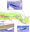
Three-dimensional reconstruction of tendons, sheath and pulleys. The SDF tendon (yellow) can be seen circumventing the DDF tendon (gold) within the synovial sheath (transparent blue). Five pulleys (red) keep the tendon closely apposed to bone. (A) Plantar view. (B) Lateral view. (C) Dorsal view. (E) Posterior oblique view. (F) Anterior oblique view. Note how the SDF forms a ring of tendon around the DDF proximal to the proximal pulley (A1).
Fig. 7
Fig. 7
Fibrocartilage regions of the DDF tendon and cellular morphology. (A) Lateral section of digit stained with alcian blue. Fibrocartilage regions indicated: 1, at level of the MCP joint; 2, in contact with the proximal pulleys; 3, in contact with the distal pulleys; and 4, overlying the middle phalanx. (B) High-magnification view of tensional tendon. Stained with Hoerscht and TRITC–phalloidin. Note the highly linear rows with cells forming contacts (arrows) with neighbouring cells. (C) High-magnification view of fibrocartilaginous tendon. Round cell morphology within lacunae can be seen (arrows).
Fig. 8
Fig. 8
Gross anatomy of the plantar vasculature of the hindpaw (plantar skin removed). (A) Tie2LacZ mouse hindpaw showing vascular endothelium (blue). (B) FITC–dextran-perfused mouse hindpaw showing perfused blood vessels (green).
Fig. 9
Fig. 9
Axial cross-sections of mouse digit vasculature. Distal to proximal sections of the digit; perfused with FITC–dextran (A–E) and endothelial LacZ expression (F–J). At the level of the insertion (A, F) there is an extensive vascular network. There is evidence of perfusate at the DDF tendon insertion (A, arrow). At the level of the DDF dorsal fibrocartilage pad numerous vascular loops can be seen in this region associated with the vincular attachment (B and G, arrows). Within the digital sheath are numerous small vascular loops (C and H, arrows). At the proximal extent of the sheath vessels can be identified at the sheath border (D and I, arrows). SDF and DDF outwith the tendon sheath (E, J). (E) The arrows show evidence of perfusate within the DDF tendon and between the SDF and DDF tendon. (J) The corresponding LacZ expression shows blood vessels between the tendons but no intratendinous LacZ expression.

Similar articles

Cited by

References

    1. Akali A, Khan U, Khaw PT, McGrouther AD. Decrease in adhesion formation by a single application of 5-fluorouracil after flexor tendon injury. Plast Reconstr Surg. 1999;103:151–158. - PubMed
    1. Anghelina M, Moldovan L, Moldovan NI. Preferential activity of Tie2 promoter in arteriolar endothelium. J Cell Mol Med. 2005;1:113–121. - PMC - PubMed
    1. Beckham C, Greenlee TK., Jr Chick vincula: elastic structures with a check-rein mechanism. J Anat. 1975;119:295–308. - PMC - PubMed
    1. Benjamin M, Ralphs JR. The cell and developmental biology of tendons and ligaments. Int Rev Cytol. 2000;196:85–130. - PubMed
    1. Chang J, Most D, Stelnicki E, et al. Gene expression of transforming growth factor beta-1 in rabbit zone II flexor tendon wound healing: evidence for dual mechanisms of repair. Plast Reconstr Surg. 1997;100:937–944. - PubMed

Publication types

LinkOut - more resources