

Microscopic and histological examination of the mouse hindpaw digit and flexor tendon arrangement with 3D reconstruction
- PMID: 17005025
- PMCID: PMC2100351
- DOI: 10.1111/j.1469-7580.2006.00625.x
Microscopic and histological examination of the mouse hindpaw digit and flexor tendon arrangement with 3D reconstruction
Abstract
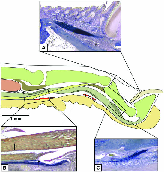
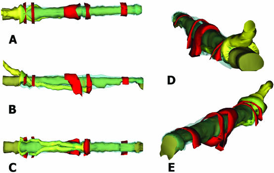
Mice are currently the species of choice for the in vivo study of injury, but few detailed anatomical descriptions have been made of rodent digits, limiting their use for the investigation of intrasynovial tendon healing. In this study a detailed microscopic and histological investigation was performed using C57/BL6 and Tie2 LacZ reporter gene transgenic mice. Serial-sectioned mouse hindpaw digits were characterized using haematoxylin and eosin, Masson's trichrome (collagen), Alcian blue (fibrocartilage), Miller's stain (elastin) and TRITC-phalloidin (cellular cytoskeleton) staining. Digital vasculature was demonstrated using FITC-labelled dextran perfusion studies supplemented with LacZ expression in Tie2 LacZ transgenic mice digits. Imaging of the digit used a combination of brightfield and confocal microscopy with three-dimensional reconstruction. Our findings demonstrated that the mouse hindpaw possesses deep and superficial flexor tendons within a synovial sheath comparable with that found in other mammalian species. The intrasynovial tendons were avascular and had regions of fibrocartilaginous specialization relating to areas of compression. Corresponding vascular networks were demonstrated around the sheath using Tie2 LacZ mice and FITC-perfused hindpaws. Furthermore, there is an area of digit where both deep and superficial tendons reside between two pulleys, similar to zone 2 in the human hand where it would be possible to study intrasynovial tendon injury and adhesion formation. In conclusion, although the dimensions of the mouse digit pose technical challenges for surgical intervention, we have identified a model for the study of flexor tendon injury that will permit future genetic manipulation studies.
Figures


References
-
- Akali A, Khan U, Khaw PT, McGrouther AD. Decrease in adhesion formation by a single application of 5-fluorouracil after flexor tendon injury. Plast Reconstr Surg. 1999;103:151–158. - PubMed
-
- Benjamin M, Ralphs JR. The cell and developmental biology of tendons and ligaments. Int Rev Cytol. 2000;196:85–130. - PubMed
-
- Chang J, Most D, Stelnicki E, et al. Gene expression of transforming growth factor beta-1 in rabbit zone II flexor tendon wound healing: evidence for dual mechanisms of repair. Plast Reconstr Surg. 1997;100:937–944. - PubMed
Publication types
MeSH terms
LinkOut - more resources
Full Text Sources
Other Literature Sources
Miscellaneous
