

Feline model of acute nipah virus infection and protection with a soluble glycoprotein-based subunit vaccine
- PMID: 17005664
- PMCID: PMC1676295
- DOI: 10.1128/JVI.01619-06
Feline model of acute nipah virus infection and protection with a soluble glycoprotein-based subunit vaccine
Abstract
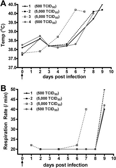
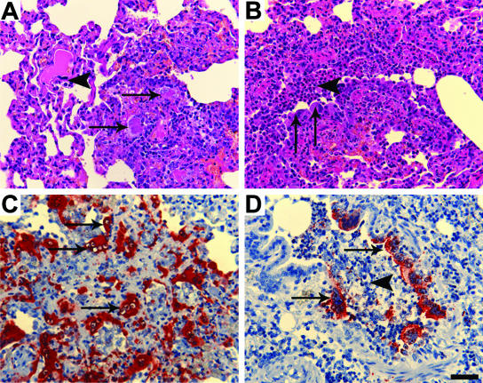
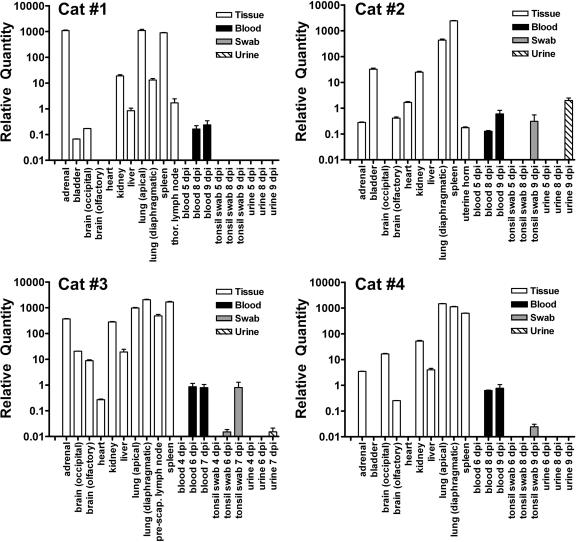
Nipah virus (NiV) and Hendra virus (HeV) are paramyxoviruses capable of causing considerable morbidity and mortality in a number of mammalian species, including humans. Case reports from outbreaks and previous challenge experiments have suggested that cats were highly susceptible to NiV infection, responding with a severe respiratory disease and systemic infection. Here we have assessed the cat as a model of experimental NiV infection and use it in the evaluation of a subunit vaccine comprised of soluble G glycoprotein (sG). Two groups of two adult cats each were inoculated subcutaneously with either 500 or 5,000 50% tissue culture infective dose(s) (TCID(50)) of NiV. Animals were monitored closely for disease onset, and extensive analysis was conducted on samples and tissues taken during infection and at necropsy to determine viral load and tissue tropism. All animals developed clinical disease 6 to 9 days postinfection, a finding consistent with previous observations. In a subsequent experiment, two cats were immunized with HeV sG and two were immunized with NiV sG. Homologous serum neutralizing titers were greater than 1:20,000, and heterologous titers were greater than 1:20,000 to 16-fold lower. Immunized animals and two additional naive controls were then challenged subcutaneously with 500 TCID(50) of NiV. Naive animals developed clinical disease 6 to 13 days postinfection, whereas none of the immunized animals showed any sign of disease. TaqMan PCR analysis of samples from naive animals revealed considerable levels of NiV genome in a wide range of tissues, whereas the genome was evident in only two immunized cats in only four samples and well below the limit of accurate detection. These results indicate that the cat provides a consistent model for acute NiV infection and associated pathogenesis and an effective subunit vaccine strategy appears achievable.
Figures
References
-
- Reference deleted.
-
- Anonymous. 2004. Nipah encephalitis outbreak over wide area of western Bangladesh, 2004. Health Sci. Bull. 2:7-11.
-
- Anonymous. 2005. Nipah virus outbreak from date palm juice. Health Sci. Bull. 3:1-5.
-
- Anonymous. 2003. Outbreaks of viral encephalitis due to Nipah/Hendra-like viruses, Western Bangladesh. Health Sci. Bull. 1:1-6.
-
- Anonymous. 2004. Person-to-person transmission of Nipah virus during outbreak in Faridpur District, 2004. Health Sci. Bull. 2:5-9.
Publication types
MeSH terms
Substances
Grants and funding
LinkOut - more resources
Full Text Sources
Other Literature Sources
Miscellaneous
