Comparative Study
. 2006 Dec;80(24):11946-59.
doi: 10.1128/JVI.01722-06. Epub 2006 Sep 27.

Evidence for CDK-dependent and CDK-independent functions of the murine gammaherpesvirus 68 v-cyclin

Affiliations
Comparative Study

Evidence for CDK-dependent and CDK-independent functions of the murine gammaherpesvirus 68 v-cyclin

Jason W Upton et al. J Virol. 2006 Dec.

Abstract

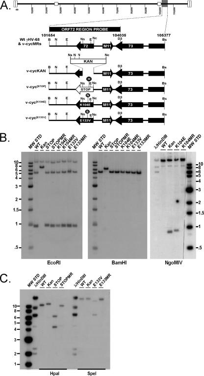
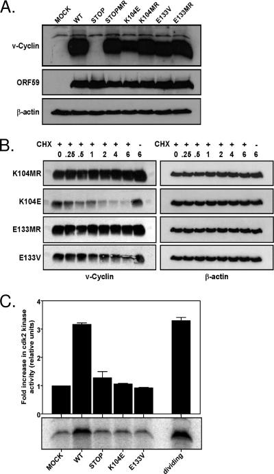
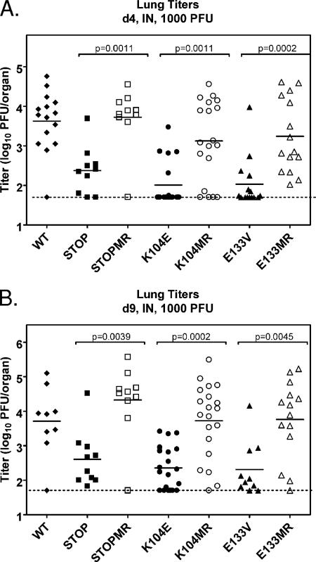
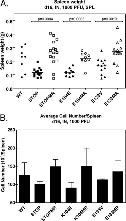
Gamma-2 herpesviruses encode homologues of mammalian D-type cyclins (v-cyclins), which likely function to manipulate the cell cycle, thereby providing a cellular environment conducive to virus replication and/or reactivation from latency. We have previously shown that the v-cyclin of murine gammaherpesvirus 68 is an oncogene that binds and activates cellular cyclin-dependent kinases (CDKs) and is required for efficient reactivation from latency. To determine the contribution of v-cyclin-mediated cell cycle regulation to the viral life cycle, recombinant viruses in which specific point mutations (E133V or K104E) were introduced into the v-cyclin open reading frame were generated, resulting in the disruption of CDK binding and activation. While in vitro growth of these mutant viruses was unaffected, lytic replication in the lungs following low-dose intranasal inoculation was attenuated for both mutants deficient in CDK binding as well as virus in which the entire v-cyclin open reading frame was disrupted by the insertion of a translation termination codon. This replication defect was not apparent in spleens of mice following intraperitoneal inoculation, suggesting a cell type- and/or route-specific dependence on v-cyclin-CDK interactions during the acute phase of virus infection. Notably, although a v-cyclin-null virus was highly attenuated for reactivation from latency, the E133V v-cyclin CDK-binding mutant exhibited only a modest defect in virus reactivation from splenocytes, and neither the E133V nor K104E v-cyclin mutants were compromised in reactivation from peritoneal exudate cells. Taken together, these data suggest that lytic replication and reactivation in vivo are differentially regulated by CDK-dependent and CDK-independent functions of v-cyclin, respectively.


Figures

FIG. 1.
FIG. 1.
Genomic structures of mutant viruses. (A) Schematic representation of γHV68 region encoding v-cyclin (ORF72) and surrounding genes from bp 101654 to bp 105377, based on the γHV68 WUMS clone sequence (73). The ORF72 region probe is depicted above the wild-type (Wt) γHV68 schematic and includes bp 101654 to bp 104035. The restriction enzyme sites shown were used for the generation of genomic clones or probes, mutagenesis, or diagnostics and are represented as follows: B, BamHI; Bs, BsrGI; D3, HinDIII; E, EcoRI; H, HpaI; N, NgoMIV; Nc, NcoI; Ns, NsiI; S, SpeI. (B and C) Southern blot analysis of wild-type (WT), mutant, and marker rescue viruses. BAC DNAs were purified, digested with the indicated restriction enzyme, electrophoresed, blotted, and hybridized with the ORF72 region probe described above. Panels labeled BamHI and EcoRI represent general diagnostic Southern analyses for all recombinants, while those labeled NgoMIV, HpaI, and SpeI are diagnostic Southern blots for the specific mutants v-cyc[K104E], v-cyc[STOP], and v-cyc[E133V], respectively. MW STD, molecular weight standard.
FIG. 2.
FIG. 2.
v-Cyclin protein expression and function. (A) Immunoblot analysis of v-cyclin protein expression. NIH 3T12 fibroblasts were lytically infected with wild-type γHV68 (WT), v-cyc[STOP], v-cyc[STOPMR],v-cyc[K104E], v-cyc[K104MR], v-cyc[E133V], or v-cyc[E133MR] or were mock infected. Cell lysates were collected at 24 h postinfection and subjected to SDS-PAGE and Western analysis. Blots were sequentially probed with rabbit polyclonal anti-v-cyclin antiserum (top panel), chicken polyclonal anti-ORF59 (middle panel), and mouse anti-β-actin (bottom panel). (B) Protein stability of mutant v-cyclin alleles. NIH 3T12 fibroblasts were lytically infected with v-cyc[K104E], v-cyc[K104MR], v-cyc[E133V], or v-cyc[E133MR]. Twenty-four hours postinfection, 50 mg/ml cycloheximide (CHX) (+) or vehicle control (−) was added to each well, and cell lysates were collected at the indicated times, in hours, posttreatment. Equivalent protein amounts were subjected to SDS-PAGE and Western analysis. Blots were probed sequentially with rabbit polyclonal anti-v-cyclin antiserum (left panels) or mouse anti-β-actin (right panels). (C) v-Cyclin mutants do not activate CDK2 activity during infection. Contact-inhibited, serum-starved NIH 3T3 fibroblasts were infected with wild-type γHV68, v-cyc[STOP], v-cyc[K104E], or v-cyc[E133V] or were mock infected in low-serum medium. Cell lysates were collected 24 h postinfection and subjected to immunoprecipitation with rabbit polyclonal anti-CDK2 directly conjugated to agarose beads. Uninfected, actively dividing NIH 3T12 fibroblasts were similarly processed as a positive control. Immunoprecipitates were used as active enzymes for in vitro kinase assays with glutathione S-transferase-pRb as an exogenous substrate. Reactions were terminated by the addition of loading buffer and boiling followed by SDS-PAGE and autoradiography. Analysis (top panel) was performed with ImageQuant software, with data normalized to mock infection. Also shown (bottom panel) is a representative experiment (one of two independent analyses).
FIG. 3.
FIG. 3.
Growth of v-cyclin mutants is similar to that of wild-type (WT) γHV68 in vitro. NIH 3T12 fibroblasts were infected with 10 PFU (A) or 0.01 PFU (B) per cell of wild-type γHV68 (closed diamonds), v-cyc[STOP] (closed squares), v-cyc[STOPMR] (open squares), v-cyc[K104E] (closed circles), v-cyc[K104MR] (open circles), v-cyc[E133V] (closed triangles), or v-cyc[E133MR] (open triangles). Inocula were removed, and samples were washed with PBS and repleted with fresh medium 1 h postinfection for determinations of single-step growth (A) or left for multistep growth analysis (B). Samples were harvested at the indicated times, and viral titers were determined by plaque assay. (C) Growth of wild-type γHV68 and v-cyclin mutants in lung epithelial cells in vitro. LA-4 lung epithelial cells were plated in low-serum medium, allowed to grow to contact inhibition, and rested for 72 h. Cells were then infected with 0.05 PFU wild-type γHV68 (closed diamonds), v-cyc[STOP] (closed squares), v-cyc[K104E] (closed circles), or v-cyc[E133V] (closed triangles) per cell in low-serum medium. Samples were harvested at the indicated times, and viral titers were determined by plaque assay. All data (A to C) are representative of two independent experiments, with titers of each sample determined in duplicate.
FIG. 4.
FIG. 4.
v-Cyclin interaction with CDKs is required for acute replication in the lung following intranasal (IN) inoculation. C57BL/6 mice were infected with 1,000 PFU of wild-type (WT) γHV68 (closed diamonds), v-cyc[STOP] (closed squares), v-cyc[STOPMR] (open squares), v-cyc[K104E] (closed circles) v-cyc[K104MR] (open circles), v-cyc[E133V] (closed triangles), or v-cyc[E133MR] (open triangles) via intranasal inoculation. Lungs were harvested at 4 (A) and 9 (B) days postinfection, and titers were determined by plaque assay. Data shown are compiled from two to three independent experiments with four to five mice each. Solid bars represent the means of each group, and the dashed line indicates the limit of detection of the assay (50 PFU/organ). Statistically significant differences (P < 0.05), as determined by nonparametric Mann-Whitney analysis, are indicated.
FIG. 5.
FIG. 5.
v-Cyclin interaction with CDKs is dispensable for acute replication in the spleen following intraperitoneal (IP) inoculation. C57BL/6 mice were infected with 1,000 PFU of wild-type (WT) γHV68 (closed diamonds), v-cyc[STOP] (closed squares), v-cyc[STOPMR] (open squares), v-cyc[K104E] (closed circles), v-cyc[K104MR] (open circles), v-cyc[E133V] (closed triangles), or v-cyc[E133MR] (open triangles) via intraperitoneal injection. Spleens were harvested at 4 (A) and 9 (B) days postinfection, and lytic titers were determined by plaque assay. Data shown are compiled from two to three independent experiments with five mice each. Solid bars represent the means of each group, and the dashed line indicates the limit of detection of the assay (50 PFU/organ). Statistically significant differences (P < 0.05) as determined by nonparametric Mann-Whitney analysis are indicated.
FIG. 6.
FIG. 6.
v-Cyclin mutant viruses do not induce splenomegaly by 16 days after intranasal infection. (A) Spleen weights of C57BL/6 mice 16 days after intranasal infection with 1,000 PFU of wild-type (WT) γHV68 (n = 8), v-cyc[STOP] (n = 15), v-cyc[STOPMR] (n = 15), v-cyc[K104E] (n = 10), v-cyc[K104MR] (n = 10), v-cyc[E133V] (n = 13), or v-cyc[E133MR] (n = 14). Statistically significant differences (P < 0.05), as determined by nonparametric Mann-Whitney analysis, are indicated. SPL, splenocytes. (B) Cell number in spleens of infected mice. Pooled samples of three to five spleens were analyzed for cell number per spleen following organ disruption and erythrocyte lysis. Data represent two to three independent experiments. Error bars represent the standard errors of the means.
FIG. 7.
FIG. 7.
Analysis of splenocyte reactivation following intranasal infection provides evidence for a CDK-independent function(s) of the γHV68 v-cyclin. C57BL/6 mice were infected with 1,000 PFU of (A) v-cyc[STOP], (B) v-cyc[K104E], (C) v-cyc[E133V], or the corresponding marker rescue virus via intranasal (IN) inoculation. Sixteen days postinfection, splenocytes (SPL) were harvested, made into single-cell suspensions, and subjected to twofold limiting dilutions on MEF indicator monolayers for determinations of the ex vivo frequencies of cells reactivating from latency. Mechanically disrupted cells were plated in parallel. Curve fit lines were derived by nonlinear regression analysis, and symbols represent means and standard errors of the means (error bars) of data from individual experiments as indicated. The dashed line (63%) represents the value used to calculate the frequency of reactivating cells as indicated by a Poisson distribution. Data represent three independent experiments, with each experiment containing cells pooled from four to five mice. (D) Graphical representation of preformed infectious virus from limiting-dilution assays (A to C). WT, wild type; CPE, cytopathic effect. Bars for each sample represent twofold dilutions of mechanically disrupted splenocytes.
FIG. 8.
FIG. 8.
Reactivation from latently infected splenocytes and PECs does not require v-cyclin-CDK interactions following intraperitoneal inoculation. C57BL/6 mice were infected with 1,000 PFU of wild-type (WT) γHV68, v-cyc[STOP], v-cyc[STOPMR], v-cyc[K104E], v-cyc[K104MR], v-cyc[E133V], or v-cyc[E133MR] via intraperitoneal (IP) injection. (A and B) Forty-two days postinfection, splenocytes (SPL) were harvested and subjected to twofold limiting dilutions on MEF indicator monolayers for determinations of the ex vivo frequency of cells reactivating from latency in the absence (A) or presence (B) of anti-IgG/IgM and anti-CD40 stimulation. Mechanically disrupted cells were plated in parallel, and preformed infectious virus was undetected in all cases (data not shown). C57BL/6 mice were infected with 1,000 PFU of wild-type γHV68 (closed diamonds), v-cyc[STOP] (closed squares), v-cyc[K104E] (closed circles), and v-cyc[E133V] (closed triangles) via intraperitoneal injection. Data represent three to four independent experiments, with each experiment containing cells pooled from four to five mice. (C) Forty-two days postinfection, PECs were harvested and subjected to twofold limiting dilutions on MEF indicator monolayers for determinations of the ex vivo frequency of cells reactivating from latency. Mechanically disrupted cells were plated in parallel, and preformed infectious virus was undetected in all cases (data not shown). Curve fit lines were derived by nonlinear regression analysis, and symbols represent means and standard errors of the means (error bars) of data from individual experiments as indicated. The dashed line (63%) represents the value used to calculate the frequency of reactivating cells as indicated by a Poisson distribution. Data represent independent experiments (wild-type γHV68, n = 6; v-cyc[STOP], n = 4; v-cyc[K104E], n = 6; v-cyc[E133V], n = 4), with each experiment containing cells pooled from four to five mice. CPE, cytopathic effect.

Similar articles

Cited by

References

    1. Adler, H., M. Messerle, and U. H. Koszinowski. 2003. Cloning of herpesviral genomes as bacterial artificial chromosomes. Rev. Med. Virol. 13:111-121. - PubMed
    1. Adler, H., M. Messerle, and U. H. Koszinowski. 2001. Virus reconstituted from infectious bacterial artificial chromosome (BAC)-cloned murine gammaherpesvirus 68 acquires wild-type properties in vivo only after excision of BAC vector sequences. J. Virol. 75:5692-5696. - PMC - PubMed
    1. Adler, H., M. Messerle, M. Wagner, and U. H. Koszinowski. 2000. Cloning and mutagenesis of the murine gammaherpesvirus 68 genome as an infectious bacterial artificial chromosome. J. Virol. 74:6964-6974. - PMC - PubMed
    1. Allen, R. D., III, S. Dickerson, and S. H. Speck. 2006. Identification of spliced gammaherpesvirus 68 LANA and v-cyclin transcripts and analysis of their expression in vivo during latent infection. J. Virol. 80:2055-2062. - PMC - PubMed
    1. Arellano, M., and S. Moreno. 1997. Regulation of CDK/cyclin complexes during the cell cycle. Int. J. Biochem. Cell Biol. 29:559-573. - PubMed

Publication types

LinkOut - more resources