. 2006 Oct;80(20):9970-6.
doi: 10.1128/JVI.01210-06.

DNA vaccination can break immunological tolerance to PrP in wild-type mice and attenuates prion disease after intracerebral challenge

Affiliations

DNA vaccination can break immunological tolerance to PrP in wild-type mice and attenuates prion disease after intracerebral challenge

Natalia Fernandez-Borges et al. J Virol. 2006 Oct.

Abstract

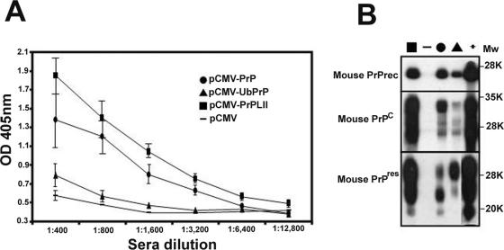
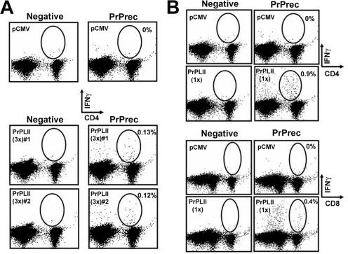
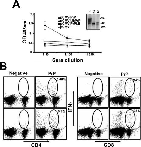
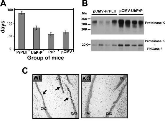
Transmissible spongiform encephalopathies (TSEs) can be ameliorated by prion protein (PrP)-specific antibodies, but active immunization is complicated by immune tolerance to the normal cellular host protein (PrP(C)). Here, we show that DNA immunization of wild-type mice can break immune tolerance against the prion protein, resulting in the induction of PrP-specific antibody and T-cell responses. PrP immunogenicity was increased by fusion to the lysosomal targeting signal from LIMPII (lysosomal integral membrane protein type II). Although mice immunized with a PrP-LIMPII DNA vaccine showed a dramatic delay in the onset of early disease signs after intracerebral challenge, immunization against PrP also had some deleterious effects. These results clearly confirm the feasibility of using active immunization to protect against TSEs and, in the absence of effective treatments, indicate a suitable alternative for combating the spread of these diseases.


Figures

FIG. 1.
FIG. 1.
DNA immunization induces PrP antibody responses. (A) Serial dilutions of sera from PrPKO mice immunized three times with pCMV (dashes), pCMV-PrP (circles), pCMV-UbPrP (triangles), and pCMV-PrPLII (squares) were tested against ELISA plates coated with recombinant prion protein. The standard deviation for each group is shown. (B) Western blot using serum from a representative mouse from each immunization group (the data were identical for all mice in the same group). All sera were diluted 1:3,200 and were tested in a Western blot assay using as antigen (i) recombinant prion protein (PrPrec; top panel), (ii) brain extracts from a WT 129/ola mouse (PrPC; middle panel), or (iii) brain extracts from a PrPSc-infected mouse after proteinase K treatment (bottom panel). As a positive control, a hyperimmune mouse serum, raised in PrP null mice against E. coli recombinant PrP protein plus Freund's adjuvant, was used at a 1:2,000 dilution (lane marked “+”). Molecular weight (Mw) standards (in thousands [K]) were used as size markers.
FIG. 2.
FIG. 2.
DNA immunization induces PrP T-cell responses. PrPKO mice were immunized three times (A) or once (B) either with pCMV or with pCMV-PrPLII. Six weeks after the final immunization, splenocytes were harvested, incubated together with the indicated semipurified protein stimulus at 1 μg/ml for 36 h, and subjected to ICCS. Circles and percentages shown (values given after subtraction of the control value) indicate the PrP-specific CD4+ T-cells (A and upper panel in B) or the PrP-specific CD8+ T-cells (B, lower panel). PrPrec, recombinant PrP.
FIG. 3.
FIG. 3.
Breaking tolerance against PrP by DNA immunization. (A) Serial dilutions of sera from WT 129/ola mice immunized three times with pCMV (dashes), pCMV-PrP (circles), pCMV-UbPrP (triangles), and pCMV-PrPLII (squares) were tested against ELISA plates coated with recombinant prion protein. The standard deviation for each group is shown. Serum from a WT 129/ola mouse immunized with pCMV-PrPLII was diluted 1:200 and tested in a Western blot assay (inset) using as the antigen (lane 1) brain extracts from a WT 129/ola mouse (PrPC), (lane 2) recombinant prion protein, or (lane 3) brain extracts from a PrPSc-infected mouse after proteinase K treatment. The data were identical for all mice in the same group. Molecular weight standards in thousands (K) were used as size markers. (B) Splenocytes from mice immunized with pCMV-PrPLII were harvested, incubated together with the indicated semipurified protein stimulus at 1 μg/ml for 36 h, and subjected to ICCS. Circles and percentages shown (values given after subtraction of the control value) indicate the PrP-specific CD4+ T cells (B, left panel) and CD8+ T cells (B, right panel) induced after DNA immunization with pCMV-PrPLII.
FIG. 4.
FIG. 4.
Vaccination confers protection against intracerebral prion challenge. (A) WT 129/ola mice were immunized three times with either pCMV, pCMV-PrP, pCMV-UbPrP, or pCMV-LII and 6 weeks later were intracerebrally challenged with PrPSc. Mice were examined weekly, and early signs of disease for each of the immunized groups were recorded and are shown. For each immunization group, all mice first developed the early symptoms within a few days of each other. (B) Mice immunized with pCMV-PrPLII or pCMV-UbPrP succumbed almost at the same time during week 22 (∼150 days) after PrPSc challenge. Equivalent amounts of brain protein extract from each dead mouse within these two vaccination groups were subjected to PK treatment (upper panel), followed by PNGase F digestion to eliminate N-linked glycans (lower panel). All four mice immunized with pCMV-PrPLII accumulated much smaller amounts of PrPres in their brains than the amounts observed in mice vaccinated with pCMV-UbPrP. (C) Twenty microliters of heat-inactivated serum from a PrPKO mouse previously immunized three times with pCMV-PrPLII (highly positive for prion-specific antibodies) was injected intracerebrally into three WT 129/ola mice or three PrPKO 129/ola mice. Twenty-four hours after intracerebral injection, mice were euthanized and brain tissues from WT mice (left panel) and PrPKO mice (right panel) were formalin fixed, cut in 5-μm-thick slices, and Nissl counterstained. Arrows indicate pyknotic neurons in the dentate gyrus (DG) and in cornu ammonis regions 2 and 3 (CA2 and CA3, respectively) of the hippocampus. Clear pyknosis can be observed in the hippocampi of WT mice (left panel), indicating neuronal death. In clear contrast, no significant neuronal damage was observed in PrPKO mice subjected to the same treatment (right panel).

References

    1. Abbott, A. 2004. Doctors seek lost data on Alzheimer's vaccine. Nature 430:715. - PubMed
    1. Aguzzi, A., and C. J. Sigurdson. 2004. Antiprion immunotherapy: to suppress or to stimulate? Nat. Rev. Immunol. 4:725-736. - PubMed
    1. Bonini, C., S. P. Lee, S. R. Riddell, and P. D. Greenberg. 2001. Targeting antigen in mature dendritic cells for simultaneous stimulation of CD4+ and CD8+ T cells. J. Immunol. 166:5250-5257. - PubMed
    1. Castilla, J., A. Gutierrez Adan, A. Brun, B. Pintado, M. A. Ramirez, B. Parra, D. Doyle, M. Rogers, F. J. Salguero, C. Sanchez, J. M. Sanchez-Vizcaino, and J. M. Torres. 2003. Early detection of PrPres in BSE infected bovine PrP transgenic mice. Arch. Virol. 148:677-691. - PubMed
    1. Castilla, J., P. Saa, C. Hetz, and C. Soto. 2005. In vitro generation of infectious scrapie prions. Cell 121:195-206. - PubMed

Publication types

MeSH terms