. 2006 Oct;80(20):9977-87.
doi: 10.1128/JVI.00354-06.

Vaccinia virus infection attenuates innate immune responses and antigen presentation by epidermal dendritic cells

Affiliations

Vaccinia virus infection attenuates innate immune responses and antigen presentation by epidermal dendritic cells

Liang Deng et al. J Virol. 2006 Oct.

Abstract

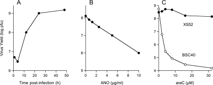
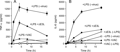
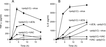
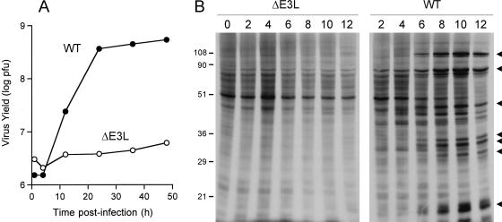
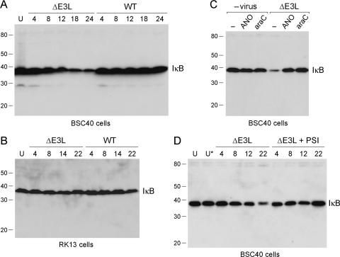
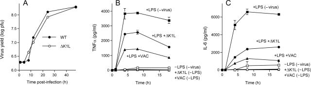
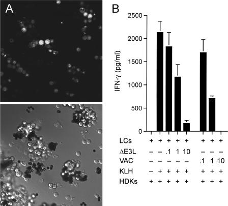
Langerhans cells (LCs) are antigen-presenting cells in the skin that play sentinel roles in host immune defense by secreting proinflammatory molecules and activating T cells. Here we studied the interaction of vaccinia virus with XS52 cells, a murine epidermis-derived dendritic cell line that serves as a surrogate model for LCs. We found that vaccinia virus productively infects XS52 cells, yet this infection displays an atypical response to anti-poxvirus agents. Whereas adenosine N1-oxide blocked virus production and viral protein synthesis during a synchronous infection, cytosine arabinoside had no effect at concentrations sufficient to prevent virus replication in BSC40 monkey kidney cells. Vaccinia virus infection of XS52 cells not only failed to elicit the production of various cytokines, including tumor necrosis factor alpha (TNF-alpha), interleukin-1beta (IL-1beta), IL-6, IL-10, IL-12 p40, alpha interferon (IFN-alpha), and IFN-gamma, it actively inhibited the production of proinflammatory cytokines TNF-alpha and IL-6 by XS52 cells in response to exogenous lipopolysaccharide (LPS) or poly(I:C). Infection with a vaccinia virus mutant lacking the E3L gene resulted in TNF-alpha secretion in the absence of applied stimuli. Infection of XS52 cells or BSC40 cells with the DeltaE3L virus, but not wild-type vaccinia virus, triggered proteolytic decay of IkappaBalpha. These results suggest a novel role for the E3L protein as an antagonist of the NF-kappaB signaling pathway. DeltaE3L-infected XS52 cells secreted higher levels of TNF-alpha and IL-6 in response to LPS and poly(I:C) than did cells infected with the wild-type virus. XS52 cells were productively infected by a vaccinia virus mutant lacking the K1L gene. DeltaK1L-infected cells secreted higher levels of TNF-alpha and IL-6 in response to LPS than wild-type virus-infected cells. Vaccinia virus infection of primary LCs harvested from mouse epidermis was nonpermissive, although a viral reporter protein was expressed in the infected LCs. Vaccinia virus infection of primary LCs strongly inhibited their capacity for antigen-specific activation of T cells. Our results highlight suppression of the skin immune response as a feature of orthopoxvirus infection.


Figures

FIG. 1.
FIG. 1.
Productive vaccinia virus infection of XS52 cells and sensitivity to antiviral agents. (A) One-step growth. XS52 cells (6 × 10⁶) were infected with vaccinia virus WR at a multiplicity of 3. The inoculum was removed after 30 min. Cells were harvested at the indicated times postinoculation. Virus yield (log PFU) was determined by titration on BSC40 cell monolayers. (B) Inhibition of virus replication by ANO. XS52 cells (1 × 10⁶) were infected at a multiplicity of 3. The inoculum was removed after 30 min and replaced with medium containing ANO at the indicated concentrations. Cells were harvested at 30 h postinfection. Virus yield (log PFU) is plotted as a function of the ANO concentration. (C) AraC blocks vaccinia virus replication in BSC40 cells but not in XS52 cells. XS52 cells (3 × 10⁶) and BSC40 cells (1 × 10⁶) were infected at a multiplicity of 3. The inocula were removed after 30 min, and the cells were overlaid with medium containing araC or control medium with no araC. Cells were harvested at 30 h postinfection. Virus yield (log PFU) is plotted as a function of the araC concentration.
FIG. 2.
FIG. 2.
ANO inhibits viral protein synthesis in XS52 cells. XS52 cells (6 × 10⁶) were pretreated with ANO (10 μg/ml) for 12 h and infected with vaccinia virus WR at a multiplicity of 10 in the presence of ANO or in the absence of the drug (Control). Cells were pulse-labeled for 30 min with [35S]methionine at 2, 4, 6, 8, and 12 h after removal of the inoculum. After the pulse, the medium was removed and the cells were lysed in situ with SDS. The labeled polypeptides were analyzed by SDS-PAGE. An autoradiograph of the gel is shown. Early viral polypeptides (indicated by filled circles) became visible at 2 to 4 h in the control infection against a background of host protein synthesis. The transition to the synthesis of late viral proteins (indicated by arrowheads) was evident at 6 to 12 h postinfection. Shutoff of host protein synthesis, also by 6 to 12 h postinfection, was evinced by decreased labeling of the prominent host polypeptide (denoted by the asterisk). The positions and sizes (in kilodaltons) of marker polypeptides are indicated on the right.
FIG. 3.
FIG. 3.
Effects of vaccinia virus infection on constitutive and LPS-induced cytokine production by XS52 cells. XS52 cells (6 × 10⁶) were infected with WT WR or ΔE3L vaccinia virus at a multiplicity of 10 or mock infected. At 2 h after removal of the inoculum, LPS was added to the medium (where indicated) to a concentration of 1 μg/ml. The concentrations of secreted TNF-α (A) and IL-6 (B) in the medium at the indicated times thereafter were determined by ELISA. Each datum represents the average of three experiments with standard deviations shown.
FIG. 4.
FIG. 4.
Effects of vaccinia virus infection on constitutive and poly(I:C)-induced cytokine production. XS52 cells (6 × 10⁶) were infected with WT WR or ΔE3L vaccinia virus at a multiplicity of 10 or mock infected. At 2 h after removal of the inoculum, poly(I:C) was added to the medium (where indicated) to a concentration of 10 μg/ml. The concentrations of secreted TNF-α (A) and IL-6 (B) in the medium at the indicated times thereafter were determined by ELISA. Each datum represents the average of three experiments with standard deviations shown.
FIG. 5.
FIG. 5.
Nonproductive infection of XS52 cells by ΔE3L. (A) One-step growth. XS52 cells were infected with WT WR or ΔE3L vaccinia virus at a multiplicity of 3. The inoculum was removed after 30 min. Cells were harvested at the indicated times postinfection. Virus yield (log PFU) is plotted as a function of time postinfection. (B) Protein synthesis in virus-infected cells. XS52 cells (6 × 10⁶) were infected with WT WR or ΔE3L vaccinia virus at a multiplicity of 10. At the indicated times postinfection, cells were pulse-labeled for 30 min with [35S]methionine as described in the legend to Fig. 2. Labeled polypeptides were analyzed by SDS-PAGE. An autoradiograph of the gel is shown. Late viral proteins are indicated by arrowheads. The positions and sizes (in kilodaltons) of marker polypeptides are indicated on the left.
FIG. 6.
FIG. 6.
Abortive ΔE3L infection triggers degradation of IκBα. (A) BSC40 cells were infected with WT WR or ΔE3L vaccinia virus at a multiplicity of 10. Whole-cell lysates were prepared from infected cells at 4, 8, 12, 18, and 24 h postinfection and also from uninfected control cells (lane U). The lysate proteins were separated by SDS-PAGE and then transferred to a membrane, which was immunoblotted with a rabbit polyclonal anti-IκBα antibody. The positions and sizes (kilodaltons) of coeloectrophoresed marker polypeptides are indicated on the left. The position of the immunoreactive IκBα protein is indicated at the right. (B) RK13 cells were infected with WT WR or ΔE3L vaccinia virus at a multiplicity of 10. Whole-cell lysates were prepared from infected cells at 4, 8, 14, and 22 h postinfection and also from uninfected control cells (lane U). The lysate proteins were separated by SDS-PAGE. A Western blot assay with rabbit polyclonal anti-IκBα antibody is shown (C). BSC40 cells were preincubated with control medium (lane −) or medium containing either ANO (10 μg/ml) or araC (8 μM) for 8 h prior to infection with ΔE3L virus at a multiplicity of 10. Virus-infected cells and mock-infected control monolayers exposed to ANO or araC were washed and incubated in fresh medium with or without ANO or araC. Whole-cell lysates were prepared at 16 h postinfection and subjected to SDS-PAGE and immunoblotting with anti-IκBα antibody. (D) BSC40 cells were infected with ΔE3L virus at a multiplicity of 10. After removal of the inoculum, the cell were overlaid with control medium or medium containing the proteasome inhibitor PSI at 0.125 μM. Lysates were prepared from ΔE3L-infected cells at 4, 8, 12, and 22 h postinfection and from uninfected cells maintained for an equivalent time in control medium (lane U) or medium containing 0.125 μM PSI (lane U*). A Western blot assay with anti-IκBα antibody is shown.
FIG. 7.
FIG. 7.
Productive replication of the ΔK1L virus in XS52 cells and effects of K1L deletion on LPS-induced cytokine production. (A) XS52 cells (6 × 10⁶) were infected with WT WR or ΔK1L vaccinia virus at a multiplicity of 3. The inoculum was removed after 30 min. Cells were harvested at the indicated times postinoculation. Virus yield (log PFU) was determined by titration on BSC40 cell monolayers. (B and C) XS52 cells (6 × 10⁶) were infected with WT WR or ΔK1L vaccinia virus at a multiplicity of 10 or mock infected. At 2 h after removal of the inoculum, LPS was added to the medium (where indicated) to a concentration of 1 μg/ml. The concentrations of secreted TNF-α (A) and IL-6 (B) in the medium at the indicated times thereafter were determined by ELISA. Each datum represents the average of three experiments with standard deviations shown.
FIG. 8.
FIG. 8.
Abortive vaccinia virus infection of primary LCs inhibits their antigen presentation function. (A) LCs were purified from female BALB/c mouse epidermis with anti-I-Ad antibody, followed by incubation with magnetic beads coated with goat anti-mouse immunoglobulin G antibody. They were infected with vaccinia virus NP-S-EGFP at a multiplicity of 10. At 24 h postinfection, the cells were imaged with a Zeiss laser scanning confocal microscope. The bright-field image is shown in the bottom half of the panel. Green fluorescence of the same field is shown in the top half of the panel. (B) Antigen presentation. Purified primary LCs were infected with vaccinia virus WR or ΔE3L at a multiplicity of 0.1, 1, or 10 or mock infected. Cells bound to magnetic beads were washed to remove unadsorbed virus, incubated overnight with the KLH antigen, and then washed to remove KLH. LCs (1 × 10⁴) were then coincubated with HDK-1 cells (5 × 10⁴) for 72 h. The IFN-γ concentration in the medium was measured by ELISA. Each datum represents the average of three antigen presentation assays with standard deviations shown.

Similar articles

Cited by

References

    1. Akira, S., S. Uematsu, and O. Takeuchi. 2006. Pathogen recognition and innate immunity. Cell 124:783-801. - PubMed
    1. Allan, R. S., C. M. Smith, G. T. Belz, A. L. van Lint, L. M. Wakim, W. R. Heath, and F. R. Carbone. 2003. Epidermal viral immunity induced by CD8α+ dendritic cells but not by Langerhans cells. Science 301:1925-1928. - PubMed
    1. Banchereau, J., and R. M. Steinman. 1998. Dendritic cells and the control of immunity. Nature 392:245-252. - PubMed
    1. Bennett, C. L., E. van Rijn, S. Jung, K. Inaba, R. M. Steinman, M. L. Kapsenberg, and B. E. Clausen. 2005. Inducible ablation of mouse Langerhans cells diminishes but fails to abrogate contact hypersensitivity. J. Cell Biol. 169:569-576. - PMC - PubMed
    1. Berthier-Vergnes, O., F. Bermond, V. Flacher, C. Massacrier, D. Schmitt, and J. Peguet-Navarro. 2005. TNF-α enhances phenotypic and functional maturation of human epidermal Langerhans cells and induces IL-12 p40 and IP-10/CXCL-10 production. FEBS Lett. 579:3660-3668. - PubMed

Publication types

MeSH terms

LinkOut - more resources