. 2006 Oct;80(20):9998-10009.
doi: 10.1128/JVI.01297-06.

Autorepression of the human cytomegalovirus major immediate-early promoter/enhancer at late times of infection is mediated by the recruitment of chromatin remodeling enzymes by IE86

Affiliations

Autorepression of the human cytomegalovirus major immediate-early promoter/enhancer at late times of infection is mediated by the recruitment of chromatin remodeling enzymes by IE86

Matthew Reeves et al. J Virol. 2006 Oct.

Abstract

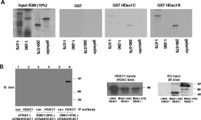
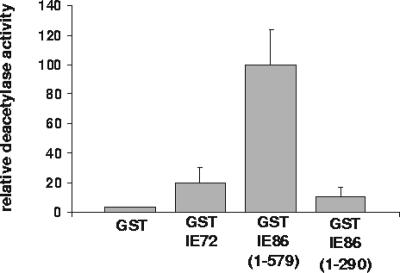
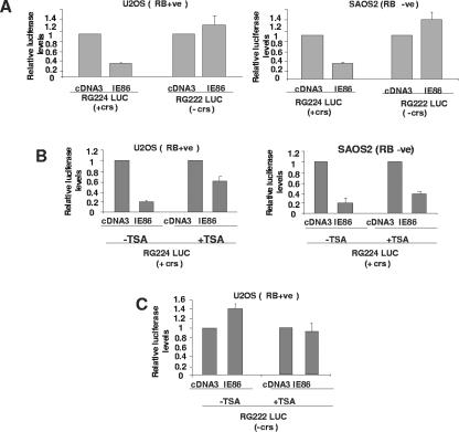
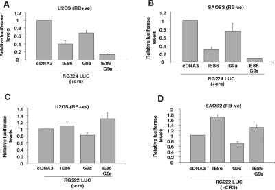
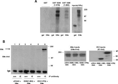
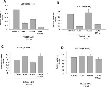
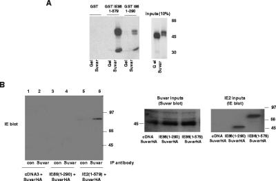
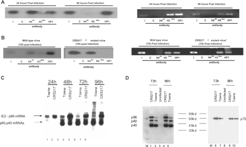
The human cytomegalovirus major immediate-early protein IE86 is pivotal for coordinated regulation of viral gene expression throughout infection. A relatively promiscuous transactivator of viral early and late gene transcription, IE86 also acts during infection to negatively regulate its own promoter via direct binding to a 14-bp palindromic IE86-binding site, the cis repression sequence (crs), located between the major immediate-early promoter (MIEP) TATA box and the start of transcription. Although such autoregulation does not involve changes in the binding of basal transcription factors to the MIEP in vitro, it does appear to involve selective inhibition of RNA polymerase II recruitment. However, how this occurs is unclear. We show that autorepression by IE86 at late times of infection correlates with changes in chromatin structure around the MIEP during the course of infection and that this is likely to result from physical and functional interactions between IE86 and chromatin remodeling enzymes normally associated with transcriptional repression of cellular promoters. Firstly, we show that IE86-mediated autorepression is inhibited by histone deacetylase inhibitors. We also show that IE86 interacts, in vitro and in vivo, with the histone deacetylase HDAC1 and histone methyltransferases G9a and Suvar(3-9)H1 and that coexpression of these chromatin remodeling enzymes with IE86 increases autorepression of the MIEP. Finally, we show that mutation of the crs in the context of the virus abrogates the transcriptionally repressive chromatin phenotype normally found around the MIEP at late times of infection, suggesting that negative autoregulation by IE86 results, at least in part, from IE86-mediated changes in chromatin structure of the viral MIEP.


Figures

FIG. 1.
FIG. 1.
IE86 interacts with HDAC1 in vitro and in vivo. (A) GST or GST fused to the C-terminal (GST HDac1C) or N-terminal (GST HDac1N) end of HDAC1 on glutathione beads was used as a target for binding to [35S]methionine-labeled full-length IE86 (1-579), a domain of IE86 containing amino acids 1 to 290 (1-290), a domain of IE86 containing amino acids 291 to 579 (291-579), or control gelsolin. Input proteins (1/10) are also shown, and molecular mass markers are in kDa. (B) SAOS2 cells were transfected with pcDNA3 (tracks 1 and 2), IE86(1-290) (tracks 3 and 4), or IE86(1-579) (tracks 5 and 6) together with pcDNA3HDAC1 expression vectors. Cell extracts were immunoprecipitated with control (con) antibody or anti-HDAC1 antibody, and immunoprecipitated complexes were separated by SDS-polyacrylamide gel electrophoresis and analyzed by Western blot analysis using an anti-IE86 antibody. Extracts were also analyzed directly for levels of expression of HDAC1 (HDAC1 inputs) by HDAC1 Western blot analysis or for expression of IE86 (IE86 inputs) by IE Western blot analysis.
FIG. 2.
FIG. 2.
IE86 has functional histone deacetylase-binding activity. GST, GST-IE72, GST-IE86(1-579), or GST-IE86(1-290) on glutathione beads was used as bait for nuclear extracts from primary human fibroblasts cells. Bound histone deacetylase activity was then assayed. The results shown are the means from triplicate samples of one experiment; the activity level of histone deacetylase binding to GST control protein was set at an arbitrary value of 1.
FIG. 3.
FIG. 3.
IE86-mediated repression of the MIEP is crs-dependent and inhibited by TSA. (A) U2OS [RB(+ve)] or SAOS2 [RB(−ve)] cells were transfected with an MIEP luciferase vector (5 μg) carrying the crs (RG224 LUC) or with a crs mutation (RG222 LUC) in the presence of 10 μg of control empty vector (cDNA3) vector or 5 μg of cDNA3IE86 (IE86) plus 5 μg of cDNA3. Cell extracts were then analyzed for luciferase activity. The results shown are the means from at least three independent experiments. (B) U2OS and SAOS2 cells were transfected and assayed as described for panel A with an MIEP luciferase vector (2.5 μg) carrying the crs (RG224 LUC) with pcDNA3 (5 μg) or pcDNA3IE86 (5 μg), but in addition, these transfections were carried out in the absence (−) or presence (+) of TSA. The results shown are the means from two independent experiments. (C) U2OS and SAOS2 cells were transfected and assayed as described for panel A but with an MIEP luciferase vector (2.5 μg) with no crs (RG222 LUC) and with pcDNA3 (5 μg) or pcDNA3IE86 (5 μg), but in addition, these transfections were carried out in the absence (−) or presence (+) of TSA. The results shown are the means from two independent experiments.
FIG. 4.
FIG. 4.
IE86 and HDAC1 act together to repress the MIEP. U2OS [RB(+ve)] (A) or SAOS2 [RB(−ve)] (B) cells were transfected with an MIEP luciferase vector carrying the crs (RG224 LUC) in the presence of 10 μg of control empty vector (cDNA3) vector, 5 μg of cDNA3IE86 (IE86) plus 5 μg of cDNA3, 5 μg of cDNA3HDAC1 (HDAC1) plus 5 μg cDNA3, or 5 μg of cDNA3IE86 plus 5 μg of cDNA3HDAC1 together. Cell extracts were then analyzed for luciferase activity. The results show the means from at least three independent experiments. The same transfections, with the exception that MIEP luciferase vector with a crs deletion (pRG222 LUC) was used, were also carried out with U2OS cells (C) and SAOS2 cells (D).
FIG. 5.
FIG. 5.
IE86 interacts with G9a in vitro and in vivo. (A) GST, GST fused to full-length IE86 (1-579), or GST fused to amino acids 1 to 290 of IE86 (1-290) on glutathione beads was used as a target for binding to [35S]methionine-labeled full-length G9a or control gelsolin (gel). Input proteins (1/10) are also shown, and molecular mass markers are in kDa. (B) SAOS2 cells were transfected with pcDNA3 (tracks 1 and 2), IE86(1-290) (tracks 3 and 4), or IE86(1-579) (tracks 5 and 6) together with G9a expression vectors. Cell extracts were immunoprecipitated with control antibody or anti-IE antibody, and immunoprecipitated complexes were separated by SDS-polyacrylamide gel electrophoresis and analyzed by Western blot analysis using a G9a antibody. Extracts were also analyzed directly for levels of expression of G9a (G9a inputs) by G9a Western blot analysis or IE86 (IE86 inputs) by IE Western blot analysis.
FIG. 6.
FIG. 6.
IE86 and G9a act together to repress the MIEP. U2OS [RB(+ve)] (A) or SAOS2 [RB(−ve)] (B) cells were transfected with an MIEP luciferase vector carrying the crs (RG224 LUC) in the presence of 10 μg of control (con) empty vector (cDNA3) vector, 5 μg of cDNA3IE86 (IE86) plus 5 μg of cDNA3, 5 μg of cDNA3G9a (G9a) plus 5 μg of cDNA3, or 5 μg of cDNA3IE86 plus 5 μg of cDNA3G9a together. Cell extracts were then analyzed for luciferase activity. The results shown are the means from at least three independent experiments. The same transfections, with the exception that the MIEP luciferase vector with a crs deletion (pRG222 LUC) was used, were also carried out with U2OS cells (C) and SAOS2 cells (D).
FIG. 7.
FIG. 7.
IE86 interacts with Suvar(3-9)H1 in vitro and in vivo. (A) GST, GST fused to full-length IE86 (1-579) or GST fused to amino acids 1 to 290 of IE86 (1-290) on glutathione beads was used as a target for binding to [35S]methionine-labeled full-length Suvar(3-9)H1 (Suvar) or control gelsolin (gel). Input proteins (1/10) are also shown, and molecular mass markers are in kDa. (B) U2OS cells were transfected with pcDNA3 (tracks 1 and 2), IE86(1-290) (tracks 3 and 4), or IE86(1-579) (tracks 5 and 6) together with Suvar(3-9)H1 expression vectors. Cell extracts were immunoprecipitated with control antibody or anti-Suvar antibody, and immunoprecipitated complexes were separated by SDS-polyacrylamide gel electrophoresis and analyzed by Western blot analysis using an E13 antibody. Extracts were also analyzed directly for levels of expression of Suvar(3-9)H1 (Suvar inputs) or IE86 (IE86 inputs) by direct Western blotting of cell extracts.
FIG. 8.
FIG. 8.
IE86 and Suvar(3-9)H1 act together to repress the MIEP, but only in the presence of RB. SAOS2 [RB(−ve)] (A) or U2OS [RB(+ve)] (B) cells were transfected with an MIEP luciferase vector carrying the crs (RG224 LUC) in the presence of 10 μg of control empty vector (cDNA3) vector, 5 μg of cDNA3IE86 (IE86) plus 5 μg of cDNA3, 5 μg of cDNA3Suvar(3-9)H1-HA plus 5 μg of cDNA3 (Suvar), or 5 μg of cDNA3IE86 plus 5 μg of cDNA3Suvar(3-9)H1-HA (IE86 Suvar) together. Cell extracts were then analyzed for luciferase activity. The results show the means from at least three independent experiments. The same transfections, with the exception that the MIEP luciferase vector with a crs deletion (pRG222 LUC) was used, were also carried out with U2OS cells (C) and SAOS2 cells (D).
FIG. 9.
FIG. 9.
The viral MIEP undergoes chromatin remodeling at late times of infection, and it is dependent on the crs. (A) Fibroblasts were infected with HCMV and analyzed by ChIPs at 24 h or 96 h postinfection using a control antibody (C), an acetylated histone H4-specific antibody (H4Ac), a dimethylated histone H3-specific antibody (H32me), or an antibody specific for HP-1. Immunocomplexes were analyzed by PCR to detect the viral MIEP. Input material (I) is also shown. The same samples were also analyzed by a PCR specific for the human γ-globin promoter (right panel). (B) Fibroblasts were infected with wild-type HCMV or HCMV with a mutated crs (CRS217 Δ) and analyzed by ChIPs at 72 h postinfection as described for panel A above. The same samples were also analyzed by a PCR specific for the human γ-globin promoter (right panel). (C) Total RNA from fibroblasts infected with Towne or CRS217 virus was isolated at 24 to 96 h postinfection, and 10 μg of the RNA was analyzed by Northern blotting. The filter was incubated with a 32P-radiolabeled exon-5-specific probe and hybridization detected by autoradiography. (D) Total protein isolated from 6 × 10³ fibroblasts infected with Towne or CRS217 virus between 72 and 96 h postinfection was analyzed by Western blot analysis for IE72 expression using an exon-4-specific antibody, and then the filter was reprobed with an anti-exon-5 antibody to show levels of IE86/p60/p40 expression.

Similar articles

Cited by

References

    1. Arlt, H., D. Lang, S. Gebert, and T. Stamminger. 1994. Identification of binding sites for the 86-kilodalton IE2 protein of human cytomegalovirus within an IE2-responsive viral early promoter. J. Virol. 68:4117-4125. - PMC - PubMed
    1. Asmar, J., L. Wiebusch, M. Truss, and C. Hagemeier. 2004. The putative zinc finger of the human cytomegalovirus IE2 86-kilodalton protein is dispensable for DNA binding and autorepression, thereby demarcating a concise core domain in the C terminus of the protein. J. Virol. 78:11853-11864. - PMC - PubMed
    1. Bannister, A. J., P. Zegerman, J. F. Partridge, E. A. Miska, J. O. Thomas, R. C. Allshire, and T. Kouzarides. 2001. Selective recognition of methylated lysine 9 on histone H3 by the HP1 chromo domain. Nature 410:120-124. - PubMed
    1. Berger, S. L. 2002. Histone modifications in transcriptional regulation. Curr. Opin. Genet. Dev. 12:142-148. - PubMed
    1. Bottardi, S., A. Aumont, F. Grosveld, and E. Milot. 2003. Developmental stage-specific epigenetic control of human beta-globin gene expression is potentiated in hematopoietic progenitor cells prior to their transcriptional activation. Blood 102:3989-3997. - PubMed

Publication types

MeSH terms

LinkOut - more resources