. 2006 Oct;80(20):10055-63.
doi: 10.1128/JVI.00985-06.

RNA silencing suppression by a second copy of the P1 serine protease of Cucumber vein yellowing ipomovirus, a member of the family Potyviridae that lacks the cysteine protease HCPro

Affiliations

RNA silencing suppression by a second copy of the P1 serine protease of Cucumber vein yellowing ipomovirus, a member of the family Potyviridae that lacks the cysteine protease HCPro

Adrian Valli et al. J Virol. 2006 Oct.

Abstract

The P1 protein of viruses of the family Potyviridae is a serine proteinase, which is highly variable in length and sequence, and its role in the virus infection cycle is not clear. One of the proposed activities of P1 is to assist HCPro, the product that viruses of the genus Potyvirus use to counteract antiviral defense mediated by RNA silencing. Indeed, an HCPro-coding region is present in all the genomes of members of the genera Potyvirus, Rymovirus, and Tritimovirus that have been sequenced. However, it was recently reported that a sequence coding for HCPro is lacking in the genome of Cucumber vein yellowing virus (CVYV), a member of the genus Ipomovirus, the fourth monopartite genus of the family. In this study, we provide further evidence that P1 enhances the activity of HCPro in members of the genus Potyvirus and show that it is duplicated in the ipomovirus CVYV. The two CVYV P1 copies are arranged in tandem, and the second copy (P1b) has RNA silencing suppression activity. CVYV P1b suppressed RNA silencing induced either by sense green fluorescent protein (GFP) mRNA or by a GFP inverted repeat RNA, indicating that CVYV P1b acts downstream of the formation of double-stranded RNA. CVYV P1b also suppressed local silencing in agroinfiltrated patches of transgenic Nicotiana benthamiana line 16c and delayed its propagation to the neighboring cells. However, neither the short-distance nor long-distance systemic spread of silencing of the GFP transgene was completely blocked by CVYV P1b. CVYV P1b and P1-HCPro from the potyvirus Plum pox virus showed very similar behaviors in all the assays carried out, suggesting that evolution has found a way to counteract RNA silencing by similar mechanisms using very different proteins in viruses of the same family.


Figures

FIG. 1.
FIG. 1.
Schematic representations of the PPV-derived (A) and CVYV-derived (B) constructs used in the RNA silencing assays. Genome maps of the viruses are shown at the top of each panel. Stop codons introduced during cloning are indicated.
FIG. 2.
FIG. 2.
Enhancement of silencing suppression activity of PPV HCPro by P1. N. benthamiana plants were coinfiltrated with A. tumefaciens carrying p35S:GFP and empty pBin19 (vector), p35S-P1PPV (P1), p35S-HCPPV (HC), p35S-P1HCPPV (P1HC), or p35S-P1PPV plus p35S-HCPPV (P1+HC). (A) GFP fluorescence pictures taken under a UV lamp at 6 dpi. (B) Northern blot analysis of GFP mRNA extracted at 6 dpi from leaf patches infiltrated with agrobacteria carrying the plasmid indicated above each lane. The bottom gel shows rRNA stained with ethidium bromide as a loading control.
FIG. 3.
FIG. 3.
Internal serine protease domain of the CVYV P1 region is functional. (A) Schematic representations of the C-terminal TAP-tagged constructs. The scissors represent serine protease domains, although processing at the end of the second one was not expected because the last residue of P1b was not included in the construct. (B) Western blot analysis of extracts of leaf patches infiltrated with agrobacteria carrying empty pBin19 (vector), p35S-P1CVYV-TAP (P1), or p35S-P1bCVYV-TAP (P1b), collected at 3 or 6 dpi. The positions of prestained molecular mass markers (New England Biolabs) (in kilodaltons) run in the same gel are indicated to the right of the gel. A band of about 45 kDa is present in samples from patches infiltrated with agrobacteria carrying p35S-P1bCVYV-TAP at 3 or 6 dpi. This minor band is recognized by the TAP antibodies and may represent a partial degradation product. The blot stained with Ponceau red is shown at the bottom as a loading control.
FIG. 4.
FIG. 4.
Suppression by CVYV P1b of RNA silencing triggered by GFP mRNA. N. benthamiana plants were coinfiltrated with agrobacteria carrying p35S:GFP and empty pBin19 (vector), p35S-P1HCPPV (P1HCPPV), p35S-P1CVYV (P1CVYV), p35S-P1aCVYV (P1aCVYV), or p35S-P1bCVYV (P1bCVYV). (A) GFP fluorescence pictures taken under a UV lamp at 6 dpi. (B) Northern blot analysis of GFP mRNA and siRNA extracted from patches infiltrated with agrobacteria carrying the plasmid indicated above each lane, collected at 3 or 6 dpi. rRNA and tRNA stained with ethidium bromide were used as loading controls for the blots of mRNA and siRNA, respectively.
FIG. 5.
FIG. 5.
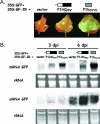
Suppression by CVYV P1b of RNA silencing triggered by GFP dsRNA. N. benthamiana plants were coinfiltrated with agrobacteria carrying plasmid p35S:GFP, p35S:GF-IR, and empty pBin19 (vector), p35S-P1HCPPV (P1HCPPV), or p35S-P1bCVYV (P1bCVYV). (A) GFP fluorescence pictures taken under a UV lamp at 6 dpi. (B) Northern blot analysis of GFP mRNA and siRNA extracted from patches infiltrated with agrobacteria carrying the plasmid indicated above each lane, collected at 3 or 6 dpi. rRNA and tRNA stained with ethidium bromide were used as loading controls for the blots of mRNA and siRNA, respectively.
FIG. 6.
FIG. 6.
Effect of CVYV P1b on systemic GFP silencing. N. benthamiana line 16c plants were coinfiltrated with agrobacteria carrying p35S:GFP and empty pBin19 (vector), p35S-P1HCPPV (P1HCPPV), or p35S-P1bCVYV (P1bCVYV). (A) GFP fluorescence pictures taken under a fluorescence microscope at 13 dpi. (B) Northern blot analysis of GFP mRNA and siRNA extracted from patches infiltrated with agrobacteria carrying the plasmid indicated above each set of pictures, collected at 3 or 7 dpi. rRNA and tRNA stained with ethidium bromide were used as loading controls for the blots of mRNA and siRNA, respectively. (C) GFP fluorescence pictures taken under a UV lamp at 13 dpi. The pictures in the bottom rows of panels B and C were taken at a magnification four times higher than that of the top rows.

Similar articles

Cited by

References

    1. Adams, M. J., J. F. Antoniw, and C. M. Fauquet. 2005. Molecular criteria for genus and species discrimination within the family Potyviridae. Arch. Virol. 150:459-479. - PubMed
    1. Anandalakshmi, R., G. J. Pruss, X. Ge, R. Marathe, A. C. Mallory, T. H. Smith, and V. B. Vance. 1998. A viral suppressor of gene silencing in plants. Proc. Natl. Acad. Sci. USA 95:13079-13084. - PMC - PubMed
    1. Baulcombe, D. 2004. RNA silencing in plants. Nature 431:356-363. - PubMed
    1. Bendahmane, A., G. Farnham, P. Moffett, and D. C. Baulcombe. 2002. Constitutive gain-of-function mutants in a nucleotide binding site-leucine rich repeat protein encoded at the Rx locus of potato. Plant J. 32:195-204. - PubMed
    1. Brigneti, G., O. Voinnet, W. X. Li, L. H. Ji, S. W. Ding, and D. C. Baulcombe. 1998. Viral pathogenicity determinants are suppressors of transgene silencing in Nicotiana benthamiana. EMBO J. 17:6739-6746. - PMC - PubMed

Publication types

MeSH terms

Associated data

LinkOut - more resources