. 2006 Oct;80(20):10117-27.
doi: 10.1128/JVI.00744-06.

The amino terminus of the herpes simplex virus 1 protein Vhs mediates membrane association and tegument incorporation

Affiliations

The amino terminus of the herpes simplex virus 1 protein Vhs mediates membrane association and tegument incorporation

Aparna Mukhopadhyay et al. J Virol. 2006 Oct.

Abstract

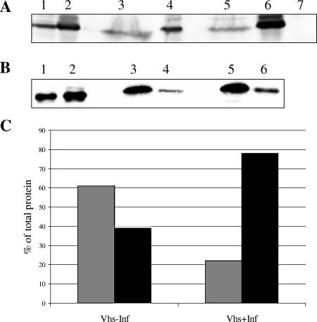
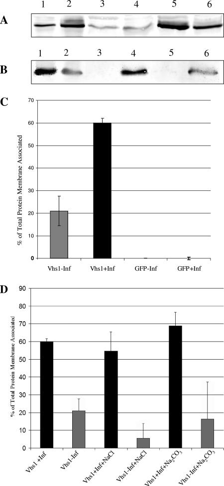
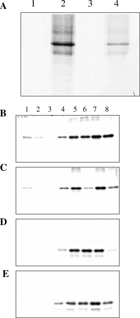
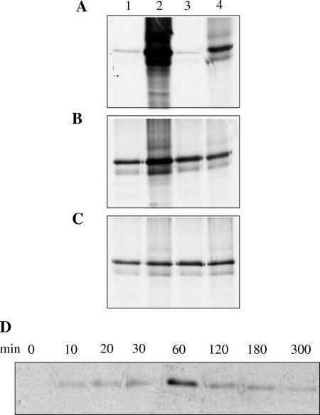
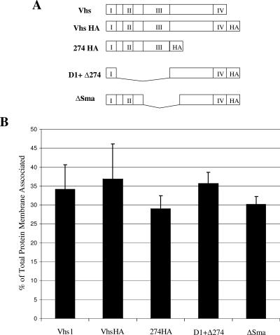
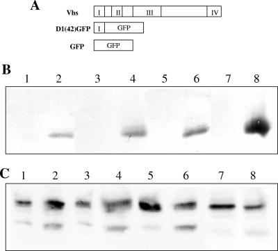
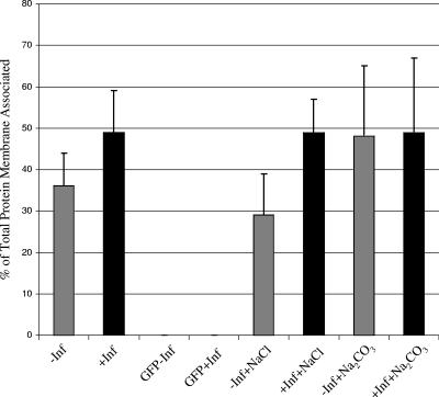
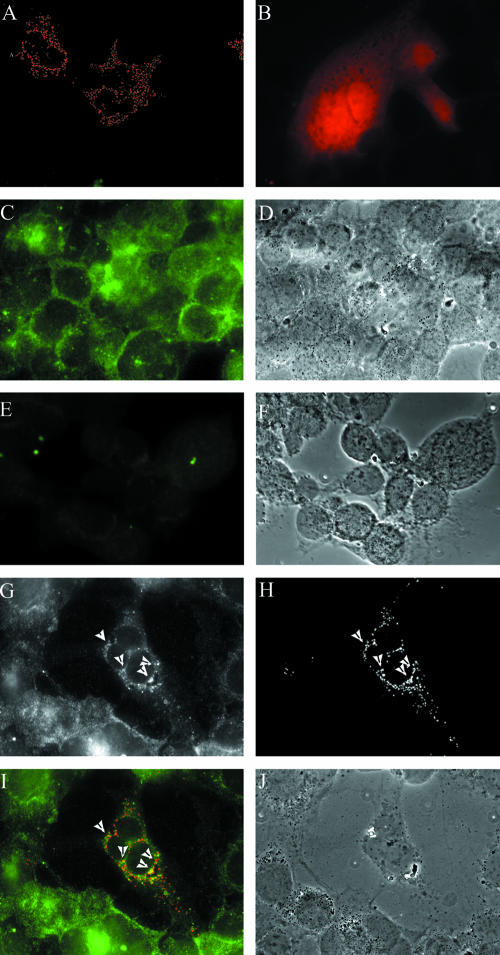
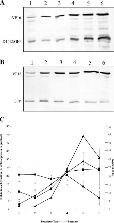
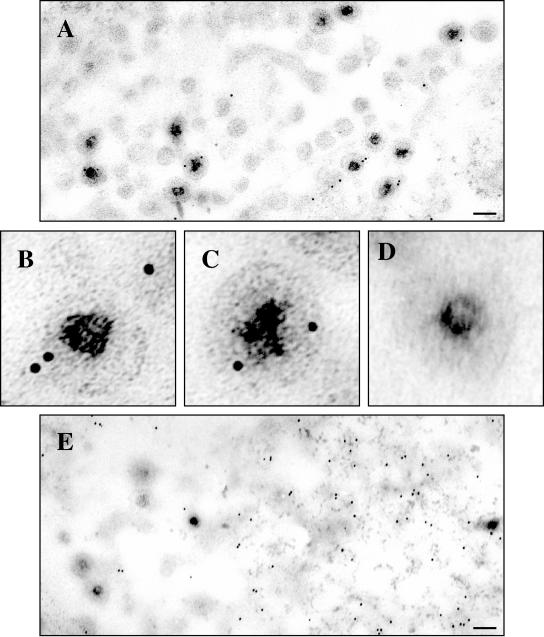
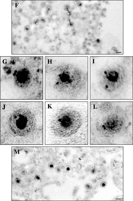
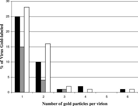
Assembly of herpes simplex viruses (HSV) is a poorly understood process involving multiple redundant interactions between large number of tegument and envelope proteins. We have previously shown (G. E. Lee, G. A. Church, and D. W. Wilson, J. Virol. 77:2038-2045, 2003) that the virion host shutoff (Vhs) tegument protein is largely insoluble in HSV-infected cells and is also stably associated with membranes. Here we demonstrate that both insolubility and stable membrane binding are stimulated during the course of an HSV infection. Furthermore, we have found that the amino-terminal 42 residues of Vhs are sufficient to mediate membrane association and tegument incorporation when fused to a green fluorescent protein (GFP) reporter. Particle incorporation correlates with sorting to cytoplasmic punctate structures that may correspond to sites of HSV assembly. We conclude that the amino terminus of Vhs mediates targeting to sites of HSV assembly and to the viral tegument.


Figures

FIG. 1.
FIG. 1.
Detergent insolubility of Vhs is largely infection dependent. COS cells were transfected with plasmids to express either Vhs1 or GFP and subsequently infected with Vhs-null virus or mock infected. The postnuclear supernatant was treated with Triton X-100 and subjected to centrifugation at 100,000 × g for 30 min. Proteins were TCA precipitated from the supernatant, and both pellet and supernatant were analyzed by Western blotting for Vhs or GFP. The distribution of (A) Vhs and (B) GFP in the detergent-soluble and -insoluble fractions is shown in the presence (lanes 2, 5, and 6) and absence (lanes 1, 3, and 4) of infection. Shown are results for total lysate (lanes 1 and 2), supernatant (lanes 3 and 5), and pellet (lanes 4 and 6). Lane 7 contained untransfected and mock-infected cell PNS demonstrating specificity of Vhs antisera. (C) Graphical representation of quantitation of the bands in panel A. Gray bars indicate supernatant, and black bars indicate pellet. −Inf, mock infected; +Inf, infected.
FIG. 2.
FIG. 2.
Membrane association of Vhs is largely infection dependent. (A and B) COS cells were transfected with plasmids to express either Vhs1 or GFP and subsequently infected with Vhs-null virus (+Inf) or mock infected (−Inf). The postnuclear supernatant was adjusted to 1.4 M sucrose and loaded at the bottom of a 1.4 M-1.2 M-0.25 M sucrose step gradient. Membranes and membrane-associated proteins at the 0.25 M-1.2 M sucrose interface and also the rest of the gradient were TCA precipitated, and analyzed by Western blotting for Vhs (A) or GFP (B). Lane 1, total lysate in absence of infection; lane 2, total lysate in the presence of infection; lane 3, membrane-associated protein in the absence of infection; lane 4, protein in the rest of the gradient in the absence of infection; lane 5, membrane-associated protein in the presence of infection; lane 6, protein in the rest of the gradient in the presence of infection. (C) Graphical representation of quantitation of the bands in panels A and B. Bands were quantitated by NIH Image J, and the amount of membrane associated is expressed as a percentage of the total. The results are the average of three independent experiments. (D) A similar experiment to those in panels A to C, but prior to gradient centrifugation, the samples were treated with either 0.5 M NaCl or 100 mM Na2CO3 as indicated. Results are the average of three independent experiments.
FIG. 3.
FIG. 3.
Membrane association of in vitro-translated Vhs. [35S]methionine-labeled Vhs1 or GFP was prepared by in vitro translation. (A) In duplicate experiments, in vitro-translated Vhs1 was subjected to sucrose density flotation and the interface fraction (lanes 1 and 3) and the rest of the gradient (lanes 2 and 4) were analyzed by SDS-PAGE. (B to E) In vitro-translated Vhs1 (B and C) or GFP (D and E) was incubated with PNS obtained from infected (B and D) and uninfected (C and E) cells and subjected to sucrose density gradient flotation. Fractions were collected and analyzed by SDS-PAGE. Lane 1 corresponds to the membrane fraction, and lane 8 is the bottom of the gradient.
FIG. 4.
FIG. 4.
Determination of optimal conditions for membrane association of in vitro-translated Vhs. (A to C) In duplicate experiments, in vitro-translated Vhs1 was incubated with infected-cell PNS for 1 h at 4°C (A), room temperature (B), and 37°C (C) before being subjected to sucrose density gradient centrifugation. The membrane fractions (lanes 1 and 3) and the rest of the gradient (lanes 2 and 4) were analyzed by SDS-PAGE. (D) In vitro-translated Vhs1 was incubated with infected-cell PNS at 37°C for various times before separation of membranes by flotation. The membrane fractions obtained were analyzed by SDS-PAGE.
FIG. 5.
FIG. 5.
Membrane association of Vhs mutants. (A) Schematic representation of Vhs1 mutants. I, II, III, and IV represent conserved domains of Vhs. (B) Graphical representation of membrane association of Vhs1 mutants in the presence of infection. The results are the average of three independent experiments.
FIG. 6.
FIG. 6.
Membrane association of D1(42)GFP in the presence and absence of infection. (A) Schematic representation of the D1(42)GFP construct used to analyze domain 1 of Vhs. (B and C) Membrane association, in duplicate, of (B) GFP or (C) D1(42)GFP in the presence (lanes 5 to 8) and absence (lanes 1 to 4) of infection. Lanes 1, 3, 5, and 7 represent the membrane fraction, whereas the corresponding rest of the gradient is in lanes 2, 4, 6, and 8.
FIG. 7.
FIG. 7.
Membrane association of D1(42)GFP after NaCl or Na2CO3 treatment. Graphical representation of D1(42)GFP membrane association in the presence (+Inf) or absence (−Inf) of infection when treated with either 0.5 M NaCl or 100 mM Na2CO3. Results are the average of two independent experiments. Lanes labeled GFP correspond to membrane association of a GFP control.
FIG. 8.
FIG. 8.
D1(42)DsRed2 colocalizes with endogenous Vhs. COS cells grown on coverslips were either transfected with D1(42)DsRed2 (A and G to J) or DsRed2 (B) or were mock transfected (C to F) and subsequently infected with virus strain N138HA (C, D, and G to J) or PAAR5 (E and F). The cells were fixed in 4% paraformaldehyde and immunostained for HA as described in Materials and Methods. The images were merged and pseudocolored using Image J software. Panels G and H are the separate green and red channel images of the same field, which has been merged in panel I. Panels D, F, and J are the phase-contrast images of panels C, E, and I, respectively.
FIG. 9.
FIG. 9.
D1(42)GFP cofractionates with extracellular virions. Extracellular virus was isolated from D1(42)GFP or GFP-transfected and infected cells by a method adapted from Szilagyi and Cunningham ( [described in Materials and Methods]). Six fractions were collected, the titer was determined for PFU, and the fractions were analyzed by Western blotting. (A and B) Western blot of VP16 and D1(42)GFP (A) and VP16 and GFP (B) in each fraction. Lane 1 represents the top of the gradient (fraction 1), and lane 6 represents the bottom (fraction 6). (C) The amount of VP16, D1(42)GFP, or GFP was quantitated in each fraction. Data from three independent experiments are plotted, representing PFU (triangles), VP16 (diamonds), D1(42)GFP (squares), and GFP (circles).
FIG.10.
FIG.10.
Immunogold electron microscopy confirms assembly of D1(42)GFP into the HSV particle. COS cells were transfected to express D1(42)GFP or GFP and subsequently infected with vhs-null virus. As a positive control, mock-transfected COS cells were infected with K26GFP virus. The cells were fixed and processed for electron microscopy as described in Materials and Methods. GFP and D1(42)GFP were detected using an anti-GFP antibody followed by a secondary antibody conjugated to 10-nm gold particles. (A to C) Cells infected with K26GFP virus. (D and E) Cells transfected with GFP and then infected. (F to M) Cells transfected with D1(42)GFP and then subsequently infected. Panel F represents a control to which no primary antibody was added. Scale bars in panels A, E, F, and M indicate 0.11 μm. Panels B to D and G to L have been enlarged fivefold relative to panels A, E, F, and M.
FIG.10.
FIG.10.
Immunogold electron microscopy confirms assembly of D1(42)GFP into the HSV particle. COS cells were transfected to express D1(42)GFP or GFP and subsequently infected with vhs-null virus. As a positive control, mock-transfected COS cells were infected with K26GFP virus. The cells were fixed and processed for electron microscopy as described in Materials and Methods. GFP and D1(42)GFP were detected using an anti-GFP antibody followed by a secondary antibody conjugated to 10-nm gold particles. (A to C) Cells infected with K26GFP virus. (D and E) Cells transfected with GFP and then infected. (F to M) Cells transfected with D1(42)GFP and then subsequently infected. Panel F represents a control to which no primary antibody was added. Scale bars in panels A, E, F, and M indicate 0.11 μm. Panels B to D and G to L have been enlarged fivefold relative to panels A, E, F, and M.
FIG. 11.
FIG. 11.
Graphical representation of percentage of viruses labeled with one to six gold particles when infection proceeded in the presence of plasmid-expressed D1(42)GFP (black bars) or GFP (gray bars) or during infection by the virus strain K26GFP (white bars). These data are from a total of 79, 114, and 129 virus particles, respectively.

Similar articles

Cited by

References

    1. Baines, J. D., R. J. Jacob, L. Simmerman, and B. Roizman. 1995. The herpes simplex virus 1 UL11 proteins are associated with cytoplasmic and nuclear membranes and with nuclear bodies of infected cells. J. Virol. 69:825-833. - PMC - PubMed
    1. Berthomme, H., B. Jacquemont, and A. Epstein. 1993. The pseudorabies virus host-shutoff homolog gene: nucleotide sequence and comparison with alphaherpesvirus protein counterparts. Virology 193:1028-1032. - PubMed
    1. Brignati, M. J., J. S. Loomis, J. W. Wills, and R. J. Courtney. 2003. Membrane association of VP22, a herpes simplex virus type 1 tegument protein. J. Virol. 77:4888-4898. - PMC - PubMed
    1. Chi, J. H., C. A. Harley, A. Mukhopadhyay, and D. W. Wilson. 2005. The cytoplasmic tail of herpes simplex virus envelope glycoprotein D binds to the tegument protein VP22 and to capsids. J. Gen. Virol. 86:253-261. - PubMed
    1. Church, G. A., and D. W. Wilson. 1997. Study of herpes simplex virus maturation during a synchronous wave of assembly. J. Virol. 71:3603-3612. - PMC - PubMed

Publication types

MeSH terms

LinkOut - more resources