. 2006 Oct;80(20):10128-38.
doi: 10.1128/JVI.00792-06.

Differential enhancement of dengue virus immune complex infectivity mediated by signaling-competent and signaling-incompetent human Fcgamma RIA (CD64) or FcgammaRIIA (CD32)

Affiliations

Differential enhancement of dengue virus immune complex infectivity mediated by signaling-competent and signaling-incompetent human Fcgamma RIA (CD64) or FcgammaRIIA (CD32)

W W Shanaka I Rodrigo et al. J Virol. 2006 Oct.

Abstract

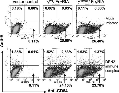
Fcgamma receptor (FcgammaR)-mediated entry of infectious dengue virus immune complexes into monocytes/macrophages is hypothesized to be a key event in the pathogenesis of complicated dengue fever. FcgammaRIA (CD64) and FcgammaRIIA (CD32), which predominate on the surface of such dengue virus-permissive cells, were compared for their influence on the infectivity of dengue 2 virus immune complexes formed with human dengue virus antibodies. A signaling immunoreceptor tyrosine-based activation motif (ITAM) incorporated into the accessory gamma-chain subunit that associates with FcgammaRIA and constitutively in FcgammaRIIA is required for phagocytosis mediated by these receptors. To determine whether FcgammaRIA and FcgammaRIIA activation functions are also required for internalization of infectious dengue virus immune complexes, we generated native and signaling-incompetent versions of each receptor by site-directed mutagenesis of ITAM tyrosine residues. Plasmids designed to express these receptors were transfected into COS-7 cells, and dengue virus replication was measured by plaque assay and flow cytometry. We found that both receptors mediated enhanced dengue virus immune complex infectivity but that FcgammaRIIA appeared to do so far more effectively. Abrogation of FcgammaRIA signaling competency, either by expression without gamma-chain or by coexpression with gamma-chain mutants, was associated with significant impairment of phagocytosis and of dengue virus immune complex infectivity. Abrogation of FcgammaRIIA signaling competency was also associated with equally impaired phagocytosis but had no discernible effect on dengue virus immune complex infectivity. These findings point to fundamental differences between FcgammaRIA and FcgammaRIIA with respect to their immune-enhancing capabilities and suggest that different mechanisms of dengue virus immune complex internalization may operate between these FcgammaRs.


Figures

FIG. 1.
FIG. 1.
Structure and expression of γ-chain/FcγRIA complex and FcγRIIA versions in COS transfectants. (A) The order of γ-chain and FcγRIA genes in the bicistronic construct ensured that FcγRIA-expressing COS cells also expressed the γ-chain (γWT/FcγRIA). An encephalomyocarditis virus-derived internal ribosomal entry site (IRES) drives the internal initiation of the FcγRIA gene. Other genes are expressed under the control of a cytomegalovirus immediate-early promoter. Stop codons inserted into the FcγRIA or γWT-chain sequence of bicistronic constructs provided control vectors. FcγRIIA was cloned into the same pcDNA5/FRT to generate a monocistronic construct (FcγRIIAWT and FcγRIIA3XMUT). A consensus Kozak sequence was introduced upstream of the γ-chain and FcγRIIA genes. Tyrosine residue positions in ITAMs of γ-chain and FcγRIIA are numbered, starting from the +1 start. (B) Solubilized lysates prepared from 2.5 × 10⁵ cells of each COS transfectant were electrophoresed and subjected to Western blotting using a monospecific rabbit serum against human γ-chain (22). (C) PE-labeled CD32 (MAb AT10) or CD64 (MAb 10.1) monoclonal antibodies and PE-labeled mouse IgG1 were used to measure the proportions of COS transfectants expressing the respective FcγR. The THP-1 human macrophage cell line served as a control. Results are representative of five or six determinations for FcγRIA transfectants and three determinations for FcγRIIA transfectants (Table 1).
FIG. 2.
FIG. 2.
Binding and phagocytosis of opsonized C. albicans by COS cells expressing FcγRIA or FcγRIIA. Rabbit IgG-sensitized, fluorescein isothiocyanate-stained yeast particles were incubated with COS cells expressing signal-competent (γWT/FcγRIA and FcγRIIAWT) or signal-incompetent (γ3XMUT/FcγRIA, γSTP/FcγRIA, and FcγRIIA3XMUT) FcγR. COS cells expressing γ-chain only or transfected with the pcDNA5/FRT vector served as controls. Phagocytosis by human macrophage-like THP-1 cells that express both FcγR was measured in parallel in each experiment. Binding and phagocytosis of opsonized C. albicans were measured using a quantitative double-fluorescence technique that employed ethidium bromide to selectively stain cell-bound (yellow) but not internalized (green) fluorescein isothiocyanate-stained yeast particles (see Materials and Methods). (A) Immunofluorescent photomicrographs (×40) of FcγR and control cells incubated with opsonized yeast particles. (B) Phagocytosis is expressed as the phagocytic index, the number of internalized yeast particles per 100 FcγR-expressing COS cells. P values: a, P < 0.02 (THP-1 versus γWT/FcγRIA or FcγRIIAWT); b, P < 0.03 (γ3XMUT/FcγRIA versus γWT/FcγRIA); c, P < 0.003 (FcγRIIA3XMUT versus FcγRIIAWT). Results are the means and standard deviations for three individual experiments with FcγRIA and four individual experiments with FcγRIIA, performed in duplicate.
FIG. 3.
FIG. 3.
Efficiencies of plaquing are comparable among COS transfectants. COS transfectants were infected with serial twofold (25 to 200) PFU dengue 2 virus (16681) in the absence of antibody. (A) Plaques were detected by indirect immunostaining with a dengue 2 virus NS1-specific monoclonal antibody. (B) The efficiencies of virus plaque formation were comparable (P = 0.17) among FcγRIA, FcγRIIA, and control (empty vector, γWT/FcγRIASTP) COS transfectants at each virus MOI. There were too many plaques to count at the 200-PFU input. Results are representative of three experiments performed in quadruplicate.
FIG. 4.
FIG. 4.
Infectivity of the virulent strain 16681 or attenuated strain New Guinea C dengue 2 virus immune complex is enhanced in COS cells that express FcγRIA or FcγRIIA. Virus-antibody complexes, prepared with serially diluted human dengue virus antiserum and dengue 2 virus (16681 [A] and NGC [B]), were added to signaling-competent (γWT/FcγRIA and FcγRIIAWT) or signaling-incompetent (γ3xMUT/FcγRIA; FcγRIIA3xMUT) Fc receptor-expressing COS cells. COS cells expressing γ-chain only (γWT/FcγRIASTP) or cells transfected with the empty pcDNA5/FRT vector served as negative controls. Plaques were detected by indirect immunostaining with a dengue 2 virus NS1-specific monoclonal antibody. Results are representative of 10 individual experiments performed in duplicate or triplicate. (C) Dengue 2 virus (16681) immune complexes were prepared by incubating virus at a single MOI (0.025) with serially diluted human dengue virus antiserum. (D) Immune complexes prepared with a single antibody dilution (1/1,000) and serial virus MOIs were added to signaling-competent (γWT/FcγRIA and FcγRIIAWT) or signaling-incompetent (γ3XMUT/FcγRIA and FcγRIIA3XMUT) Fc receptor-expressing COS cells. Cells expressing γ-chain only (γWT/FcγRIASTP) or those transfected with the pcDNA5/FRT vector served as negative controls. Plaques were detected by indirect immunostaining with a dengue 2 virus NS1-specific monoclonal antibody. For FcγRIIA, plaques corresponding to an MOI of 0.5 were too numerous to count. P values were determined using a two-tailed t test. a, γWT/FcγRIA versus γ3XMUT/FcγRIA; b, γ3XMUT/FcγRIA versus γWT/FcγRIASTP and vector controls. For panel C: a, P < 0.001; b, P < 0.03. For panel D: a, P < 0.005; b, P < 0.007. Results are the means and standard deviations for an experiment performed in quadruplicate and are representative of three individual experiments performed in triplicate or quadruplicate.
FIG. 5.
FIG. 5.
Infection of signaling-competent and -incompetent, FcγRIA-expressing COS cells measured by flow cytometry. COS cells expressing γWT/FcγRIA or γ3XMUT/FcγRIA and control vector COS transfectants were infected with preformed dengue virus immune complexes (MOI, 0.25; dengue virus antiserum dilution, 1/4,000) or mock infected and analyzed by flow cytometry 2 days postinfection with Alexa 647-labeled anti-dengue 2 virus envelope E protein (7E1) or PE-labeled anti-CD64 monoclonal antibodies, respectively.
FIG. 6.
FIG. 6.
FcγRIA bereft of γ-chain mediates dengue virus immune enhancement. Dengue 2 virus (16681) immune complexes prepared by incubating virus at a single MOI (0.25) and human dengue virus antiserum dilution (1/500 to 1/4,000 [A] and 1/4,000 [B]) were incubated in cluster plates overnight with trypsinized COS cells expressing FcγRIA alone (γSTP/FcγRIA) or in association with native (γWT/FcγRIA) or signaling-incompetent γ-chain (γ3XMUT/FcγRIA). After overnight incubation, an agarose overlay was added to the monolayered cells (A) or monolayers were washed to remove residual virus and replenished with fresh medium (B). In the experiment whose results are shown in panel A, agarose plugs were removed on the second postinfection day and plaques were detected by indirect immunostaining with a dengue 2 virus NS1-specific monoclonal antibody. Each condition was tested in quadruplicate. a, P = 0.06 (γ3XMUT/FcγRIA versus γSTP/FcγRIA); b, P < 0.001 (γSTP/FcγRIA versus vector control). In the experiment whose results are shown in panel B, supernatants were removed in their entirety from the respective wells daily and dengue virus was titrated by plaque assay in Vero cells. Virus concentrations are expressed as the log10 means ± standard deviations for an experiment performed in triplicate and titrated in duplicate. P < 0.003 (vector control versus γWT/FcγRIA, γ3XMUT/FcγRIA, and γSTP/FcγRIA); P < 0.015 (γWT/FcγRIA versus γ3XMUT/FcγRIA or γSTP/FcγRIA).
FIG. 7.
FIG. 7.
FcγRIA-mediated phagocytosis and dengue virus immune complex infectivity are proportionately reduced by selective γ-chain mutation. COS cells were transfected with bicistronic vectors composed of γ-chain alone (γWT/FcγRIASTP), FcγRIA alone (γSTP/FcγRIA), or FcγRIA and γ-chain, in which its cytoplasmic tail residues (Y58, Y65, and Y76) were individually or multiply (γY65,76 and γ3XMUT) mutated by Tyr-to-Phe residue substitution. Results are from an experiment performed in triplicate. (A) γ-Chain abundance determined by Western blotting. Solubilized lysates prepared from 2.5 × 10⁵ cells of each COS transfectant were electrophoresed and subjected to Western blotting using a monospecific rabbit serum against human γ-chain (22). (B) Phagocytoses of opsonized yeast particles and infectivities of dengue virus immune complexes. COS cells were transfected with γ-chain/FcγRIA versions or FcγRIA only. COS cells transfected with an empty vector or expressing γ-chain only (γWT/FcγRIASTP) served as controls. The phagocytic index was defined as the number of yeast particles internalized by 100 FcγRIA-expressing COS cells. In parallel, the respective COS transfectants were incubated with dengue 2 virus (16681) immune complexes formed with pooled human dengue virus antiserum (1/1,000) and serial concentrations of dengue virus; results for an experiment performed in triplicate with a virus MOI of 1.0 are shown. For both assays, the results were normalized against those for the γWT/FcγRIA transfectant. All γ-chain ITAM mutations and the γ-chain deletion (γSTP/FcγRIA) led to significant (P < 0.05, two-tailed t test) reductions in immune complex infectivity compared to that observed with the γWT/FcγRIA transfectant. (C) Correlation between phagocytosis and infectivity among dengue virus immune complexes formed with dengue virus antisera (1/1,000 dilution) and dengue 2 virus with MOIs of 0.25 (a), 0.50 (b), and 1.0 (c).

References

    1. Aderem, A., and D. M. Underhill. 1999. Mechanisms of phagocytosis in macrophages. Annu. Rev. Immunol. 17:593-623. - PubMed
    1. Almeida, J. D., and A. P. Waterson. 1969. The morphology of virus-antibody interaction. Adv. Virus Res. 15:307-338. - PMC - PubMed
    1. Daeron, M. 1997. Fc receptor biology. Annu. Rev. Immunol. 15:203-234. - PubMed
    1. Davis, W., P. T. Harrison, M. J. Hutchinson, and J. M. Allen. 1995. Two distinct regions of FC gamma RI initiate separate signalling pathways involved in endocytosis and phagocytosis. EMBO J. 14:432-441. - PMC - PubMed
    1. Duchemin, A. M., L. K. Ernst, and C. L. Anderson. 1994. Clustering of the high affinity Fc receptor for immunoglobulin G (Fc gamma RI) results in phosphorylation of its associated gamma-chain. J. Biol. Chem. 269:12111-12117. - PubMed

Publication types

LinkOut - more resources