Comparative Study
. 2006 Oct;80(20):10139-50.
doi: 10.1128/JVI.00854-06.

EBNA-3B- and EBNA-3C-regulated cellular genes in Epstein-Barr virus-immortalized lymphoblastoid cell lines

Affiliations
Comparative Study

EBNA-3B- and EBNA-3C-regulated cellular genes in Epstein-Barr virus-immortalized lymphoblastoid cell lines

Adrienne Chen et al. J Virol. 2006 Oct.

Abstract

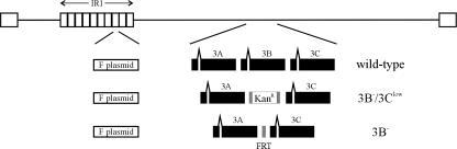
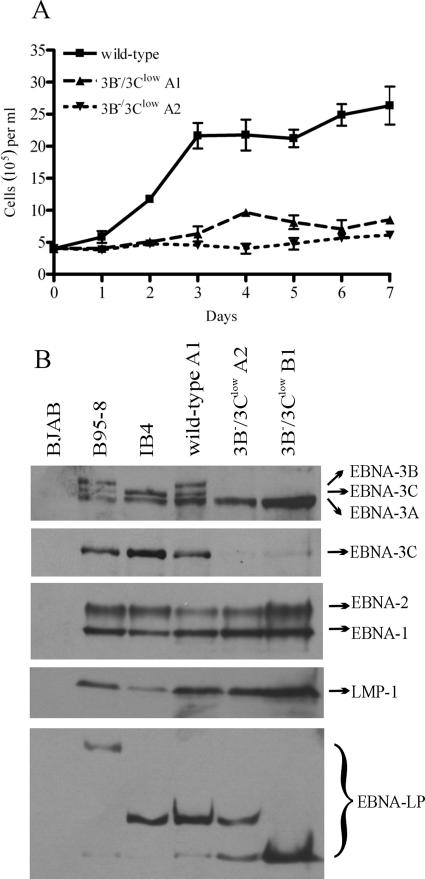
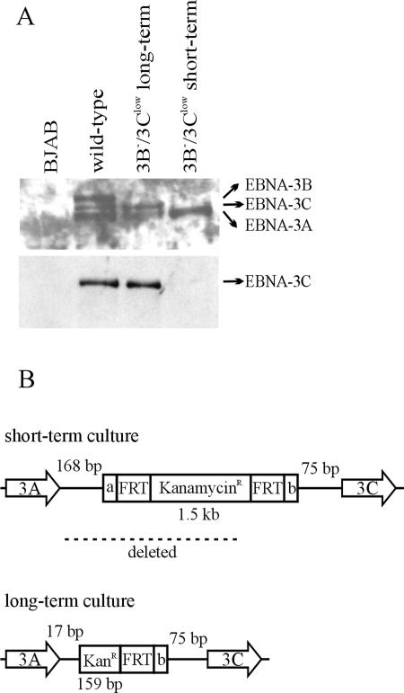
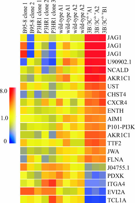
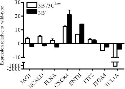
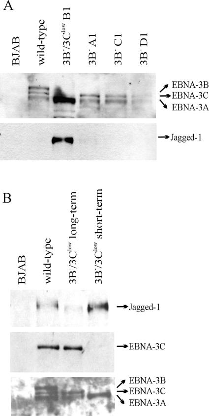
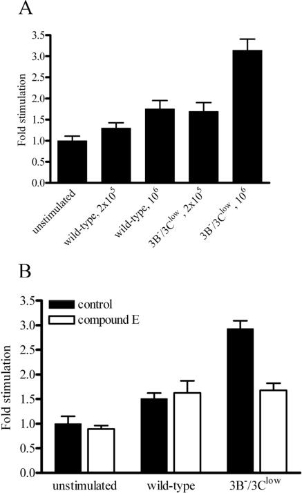
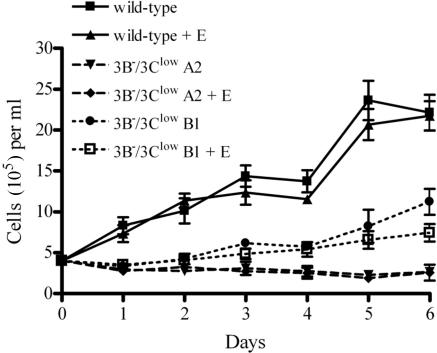
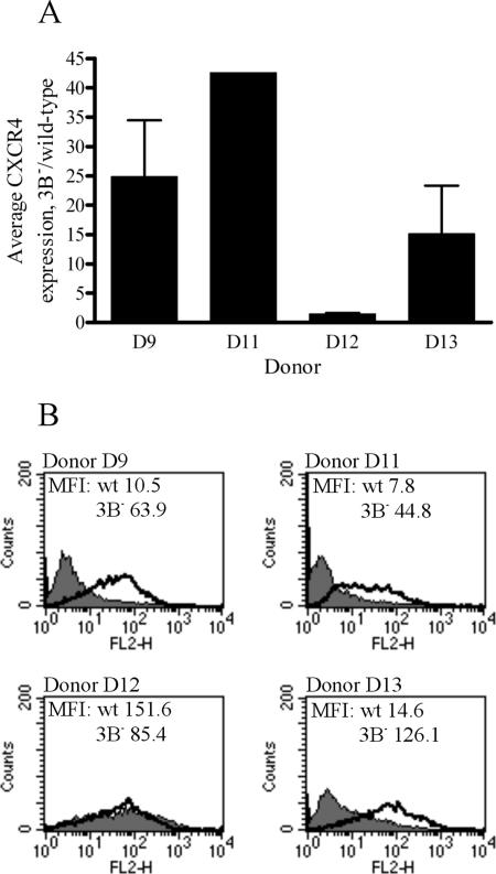
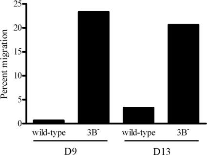
The cellular pathways that Epstein-Barr virus (EBV) manipulates in order to effect its lifelong persistence within hosts and facilitate its transmission between hosts are not well understood. The EBV nuclear antigen 3 (EBNA-3) family of latent infection proteins consists of transcriptional regulators that influence viral and cellular gene expression in EBV-infected cells. To identify EBNA-3B- and EBNA-3C-regulated cellular genes potentially important for virus infection in vivo, we studied a lymphoblastoid cell line (LCL) infected with an unusual EBV mutant, where a genetic manipulation to delete EBNA-3B also resulted in a significant decrease in EBNA-3C expression and slower than normal growth (3B(-)/3C(low)). Transcriptional profiling was performed on the 3B(-)/3C(low) LCLs, and comparison of mutant and wild-type LCL profiles resulted in a group of 21 probe sets representing 16 individual genes showing statistically significant differences in expression. Further quantitative reverse transcription-PCR analyses comparing 3B(-)/3C(low) LCLs to a previously described EBNA-3B mutant (3B(-)) where EBNA-3C expression was normal revealed three potential EBNA-3B-repressed genes, three potential EBNA-3C-repressed genes, and two potential EBNA-3C-activated genes. The most highly EBNA-3C-repressed gene was Jagged1, a cell surface ligand and inducer of the Notch receptor signaling pathway that is usurped by EBV genes essential for B-cell immortalization. 3B(-)/3C(low) LCLs expressed increased levels of Jagged1 protein and were able to more efficiently induce functional Notch signaling, and this signaling was dependent on Notch cleavage by gamma-secretase. However, inhibiting gamma-secretase-mediated Notch cleavage did not rescue 3B(-)/3C(low) LCL growth, suggesting that EBNA-3C-mediated repression of this signaling pathway did not contribute to LCL growth in tissue culture. Similarly, expression of the chemokine receptor CXCR4 was reproducibly upregulated in EBNA-3B-null LCLs. Since deletion of EBNA-3B has no significant impact on B-cell immortalization in tissue culture, this finding suggested that EBNA-3B-mediated regulation of CXCR4 may be an important viral strategy for alteration of B-cell homing in the infected host. These studies identify two cellular genes that do not contribute to EBV-induced B-cell growth but whose expression levels are strongly EBNA-3 regulated in EBV-infected primary B cells. These EBV-manipulated cellular pathways may be important for virus survival or transmission in humans, and their independence from EBV-induced B-cell growth makes them potential targets for testing in vivo with the rhesus lymphocryptovirus animal model for EBV infection.


Figures

FIG. 1.
FIG. 1.
Schematic of EBNA-3B mutations made in the EBV BAC. F plasmid sequences for prokaryotic replication were inserted into the EBV internal repeat 1 (IR1) region. The wild-type BAC contains the full complement of EBNA-3A, -3B, and -3C genes. EBNA-3B was knocked out in the 3B/3Clow BAC by replacement of the entire EBNA-3B open reading frame with a kanamycin resistance cassette (Kanr) flanked by FRT sites. The 3B BAC has been described previously and contains a single FRT site in place of the EBNA-3B open reading frame (2).
FIG. 2.
FIG. 2.
3B/3Clow LCLs are impaired for cell growth and express low levels of EBNA-3C protein. (A) Growth curves for wild-type and 3B/3Clow LCLs. Wild-type and two independently derived 3B/3Clow LCLs (3B/3Clow A1 and A2) were seeded at an initial density of 4 × 10⁵ cells per ml in a 24-well plate at day 0, and viable cell counts were taken over a period of 7 days. Data are means ± standard deviations for three replicates. (B) Expression of EBV latent proteins in 3B/3Clow LCLs. Whole-cell lysates were analyzed for expression of EBV latent proteins by Western blotting. EBV-immune human serum was used to detect EBNA-1, -2, -3A, -3B, and -3C; EBNA-3C alone was detected using the A10 monoclonal antibody (30); EBNA-LP was detected using the JF186 monoclonal antibody (9); and LMP-1 was detected using the S12 monoclonal antibody (27). Controls included an EBV-negative Burkitt lymphoma line (BJAB), an LCL infected with the B95-8 virus (B95-8), and an LCL derived from B95-8-immortalized umbilical-cord B cells (IB4) (21) which lacks full-length EBNA-3B protein expression (38) but is normal for expression of all the other latent proteins. An LCL infected with the wild-type BAC-derived virus (wild-type) and two independently derived 3B/3Clow LCLs are shown.
FIG. 3.
FIG. 3.
Spontaneous restoration of normal EBNA-3C expression in a 3B/3Clow LCL after several months in culture. (A) Expression of the EBNA-3 proteins in a 3B/3Clow LCL after prolonged culture. Whole-cell lysates were analyzed for expression of EBNA-3 proteins by Western blotting. EBV-immune serum was used to detect all the EBNA-3s, and the A10 monoclonal antibody was used to detect EBNA-3C alone. (B) Schematic of the mutation in the EBNA-3C restored 3B/3Clow LCL. The DNA region between the EBNA-3A and EBNA-3C open reading frames was PCR amplified and sequenced. KanamycinR/KanR represents the kanamycin resistance cassette inserted in the place of the EBNA-3B ORF; FRT represents the FLP recombinase target sites flanking the Kanr cassette; a and b represent nonviral, unique flanking sequences on either side of the FRT sites. The numbers of EBV nucleotides present between the nonviral insert and the EBNA-3A stop codon or EBNA-3C start codon are indicated by the numbered base pairs (bp).
FIG. 4.
FIG. 4.
Altered cellular gene expression in 3B/3Clow BAC-infected LCLs. Shown are genes displaying significant changes in expression levels in 3B/3Clow LCLs relative to expression levels in all wild-type LCLs grouped together (B95-8, P3HR1, and wild-type BAC-derived LCLs). Vertical columns represent data obtained for each individual cell line: for two LCLs immortalized with B95-8 virus (B95-8 clones 1 and 2), three LCLs derived by homologous recombination of EBNA-LP and EBNA-2 in P3HR1 EBV (P3HR1 clones 1, 2, and 3), two LCLs derived from the wild-type EBV BAC (wild-type A1 and A2), and three LCLs derived from the 3B/3Clow BAC (3B/3Clow A1, A2, and B1). Horizontal rows represent data obtained for each individual gene across all cell lines. The color of each data point represents the normalized signal-to-control ratio for a given gene in a specific cell line. Identities of individual genes are as follows: JAG1, jagged1; NCALD, neurocalcin D; AKR1C1, aldoketoreductase family, member C1; UST, uronyl-2-sulfotransferase; CHST4, carbohydrate sulfotransferase 4; CXCR4, chemokine (C-X-C motif) receptor 4; ENTH, enthoprotin; AIM1, absent in melanoma 1; P101-PI3K, phosphoinositide-3-kinase, regulatory subunit, polypeptide p101; TTF2, transcription termination factor, RNA polymerase II; JWA, vitamin A responsive; FLNA, filamin A, alpha; PDXK, pyridoxal kinase; ITGA4, integrin α4; EVI2A, ecotropic viral integration site 2A; TCL1A, T-cell leukemia/lymphoma 1A. GenBank accession numbers for the two unknown genes are shown.
FIG. 5.
FIG. 5.
Quantitative RT-PCR analysis of cellular gene expression levels in 3B/3Clow and 3B BAC-derived LCLs. Quantitative RT-PCR was performed on RNA derived from wild-type, 3B/3Clow, and 3B LCLs. Average CT values for each gene were normalized to GAPDH levels, and data displayed are changes (n-fold) in expression levels of 3B/3Clow and 3B LCLs relative to expression levels in wild-type LCLs. Data are means ± standard deviations for two independent experiments performed in triplicate on the same RNA preparation.
FIG. 6.
FIG. 6.
Jagged1 protein expression is increased in 3B/3Clow BAC-derived LCLs. (A) Whole-cell lysates from wild-type, 3B/3Clow, and 3B BAC-derived LCLs were analyzed for EBNA-3 and Jagged1 expression by Western blotting. EBV-immune human serum was used to detect the EBNA-3 proteins; Jagged1 protein was detected using the H-114 polyclonal antibody. (B) Downregulation of Jagged1 expression in 3B/3Clow LCLs that are restored for EBNA-3C expression. Whole-cell lysates from the 3B/3Clow LCL restored for normal EBNA-3C expression were analyzed for Jagged1 expression by Western blotting. The bottom two panels are reproduced from Fig. 3A, for reference and loading control purposes.
FIG. 7.
FIG. 7.
3B/3Clow LCLs are able to induce Notch signaling. (A) Notch-induced luciferase activity in U2OS cells stimulated with wild-type or 3B/3Clow LCLs. U2OS cells were transfected with a Notch luciferase reporter construct containing four RBP-Jκ binding sites in its promoter (17). Twenty-four hours posttransfection, cells were overlaid with either wild-type or 3B/3Clow LCLs at different concentrations. Forty-eight hours posttransfection, cells were harvested and lysates assayed for luciferase activity. Data shown are stimulation levels (n-fold) relative to those in unstimulated controls and represent the means ± standard deviations for three replicates. (B) Inhibition of 3B/3Clow LCL-stimulated, Notch-induced luciferase activity by the γ-secretase inhibitor compound E. U2OS cells were transfected with luciferase reporter constructs and treated with 1 μM compound E. Twenty-four hours posttransfection, cells were overlaid with either wild-type or 3B/3Clow LCLs at a density of 1 × 10⁶ cells/well. Forty-eight hours posttransfection, cells were harvested and lysates assayed for luciferase activity. Data shown are stimulation levels (n-fold) relative to those in untreated, unstimulated controls and represent the means ± standard deviations for three replicates.
FIG. 8.
FIG. 8.
Notch signaling induced by 3B/3Clow BAC-derived LCLs is not responsible for their slow growth. Wild-type and 3B/3Clow LCLs (3B/3Clow A1 and 3B/3Clow B1) were treated with either dimethyl sulfoxide or 1 μM compound E (+E) at day 0. Cells were seeded at an initial density of 4 × 10⁵ cells per ml in a 24-well plate at day 0, and cell counts were taken over a period of 6 days. Data shown are means ± standard deviations for three replicates.
FIG. 9.
FIG. 9.
RNA and cell surface protein expression of CXCR4 in 3B LCLs from multiple donors. (A) Average expression levels of CXCR4 mRNA in 3B LCLs, relative to expression levels in wild-type LCLs. Multiple independent wild-type and 3B LCLs were generated from four different donors, and quantitative RT-PCR for CXCR4 was performed on RNA isolated from these cells. Average CT values for CXCR4 were normalized to GAPDH levels, and data displayed are changes (n-fold) in expression levels in 3B LCLs relative to expression levels in wild-type LCLs. Data are means ± standard deviations for independent experiments performed in triplicate on independently derived 3B LCLs. (B) Cell surface expression of CXCR4 protein in wild-type (wt) and 3B LCLs from different donors. Wild-type and 3B LCLs were stained for CXCR4 expression by using a PE-conjugated antibody directed against CXCR4 (clone 1D9). A PE-conjugated, isotype-matched antibody was used as a control. The shaded area shows CXCR4 staining of wild-type LCLs, and the bold line represents staining of 3B LCLs. The mean fluorescence intensities (MFI) of each sample are indicated on their respective graphs.
FIG. 10.
FIG. 10.
Increased CXCR4 expression on 3B LCLs restores their ability to migrate in response to ligand. Wild-type and 3B LCLs from two different donors (D9 and D13) were loaded into the upper chamber of a Transwell insert, and their chemotactic responses to 100 ng/ml CXCL12 (SDF-1α) were compared after 4 h. Percent migration was calculated as the fraction of total cells migrating across the Transwell filter, after subtracting the number of cells migrating in response to medium alone.

Similar articles

Cited by

References

    1. Babcock, G. J., L. L. Decker, M. Volk, and D. A. Thorley-Lawson. 1998. EBV persistence in memory B cells in vivo. Immunity 9:395-404. - PubMed
    1. Chen, A., M. Divisconte, X. Jiang, C. Quink, and F. Wang. 2005. Epstein-Barr virus with the latent infection nuclear antigen 3B completely deleted is still competent for B-cell growth transformation in vitro. J. Virol. 79:4506-4509. - PMC - PubMed
    1. Cludts, I., and P. J. Farrell. 1998. Multiple functions within the Epstein-Barr virus EBNA-3A protein. 72:1862-1869. - PMC - PubMed
    1. Cotter, M. A., II, and E. S. Robertson. 2000. Modulation of histone acetyltransferase activity through interaction of Epstein-Barr nuclear antigen 3C with prothymosin alpha. Mol. Cell. Biol. 20:5722-5735. - PMC - PubMed
    1. D'Apuzzo, M., A. Rolink, M. Loetscher, J. A. Hoxie, I. Clark-Lewis, F. Melchers, M. Baggiolini, and B. Moser. 1997. The chemokine SDF-1, stromal cell-derived factor 1, attracts early stage B cell precursors via the chemokine receptor CXCR4. Eur. J. Immunol. 27:1788-1793. - PubMed

Publication types

MeSH terms

LinkOut - more resources