. 2006 Oct;80(20):10162-72.
doi: 10.1128/JVI.00249-06.

Infection of CD127+ (interleukin-7 receptor+) CD4+ cells and overexpression of CTLA-4 are linked to loss of antigen-specific CD4 T cells during primary human immunodeficiency virus type 1 infection

Affiliations

Infection of CD127+ (interleukin-7 receptor+) CD4+ cells and overexpression of CTLA-4 are linked to loss of antigen-specific CD4 T cells during primary human immunodeficiency virus type 1 infection

John J Zaunders et al. J Virol. 2006 Oct.

Abstract

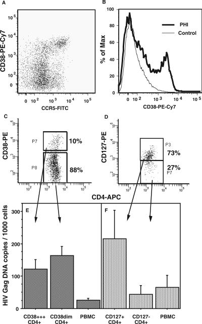
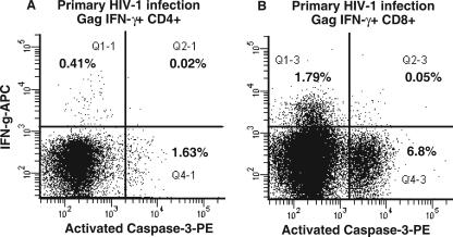
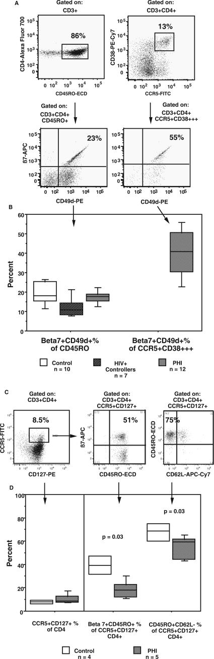
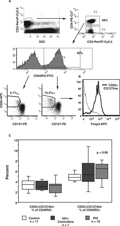
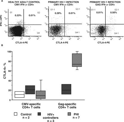
We recently found that human immunodeficiency virus (HIV)-specific CD4+ T cells express coreceptor CCR5 and activation antigen CD38 during early primary HIV-1 infection (PHI) but then rapidly disappear from the circulation. This cell loss may be due to susceptibility to infection with HIV-1 but could also be due to inappropriate apoptosis, an expansion of T regulatory cells, trafficking out of the circulation, or dysfunction. We purified CD38+++CD4+ T cells from peripheral blood mononuclear cells, measured their level of HIV-1 DNA by PCR, and found that about 10% of this population was infected. However, a small subset of HIV-specific CD4+) T cells also expressed CD127, a marker of long-term memory cells. Purified CD127+CD4+ lymphocytes contained fivefold more copies of HIV-1 DNA per cell than did CD127-negative CD4+ cells, suggesting preferential infection of long-term memory cells. We observed no apoptosis of antigen-specific CD4+ T cells in vitro and only a small increase in CD45RO+CD25+CD127dimCD4+ T regulatory cells during PHI. However, 40% of CCR5+CD38+++ CD4+ T cells expressed gut-homing integrins, suggesting trafficking through gut-associated lymphoid tissue (GALT). Furthermore, 80% of HIV-specific CD4+ T cells expressed high levels of the negative regulator CTLA-4 in response to antigen stimulation in vitro, which was probably contributing to their inability to produce interleukin-2 and proliferate. Taken together, the loss of HIV-specific CD4+ T cells is associated with a combination of an infection of CCR5+ CD127+ memory CD4+ T cells, possibly in GALT, and a high expression of the inhibitory receptor CTLA-4.


Figures

FIG. 1.
FIG. 1.
HIV DNA in CD38+++ and CD127+ subpopulations of CD4+ cells from PBMCs during PHI. A representative dot plot of CCR5+CD38+++ CD4+ T cells present in fresh whole blood during PHI is shown in panel A, and a representative comparison of CD38 expression on CD4+ T cells between a subject during PHI and a healthy adult control subject is shown in panel B. PBMCs were stained with CD4-APC and CD38-PE (C) or CD4-APC and CD127-PE (D) and sorted into different subpopulations. Representative histograms for each of the two sorting protocols are shown. DNA was extracted from purified cells, and the number of copies of HIV DNA per cell in each subpopulation was determined by quantitative PCR and normalized to beta-actin. Each column shows the mean (and standard error) from three independent experiments for each subpopulation of CD4+ cells defined by CD38 (E) or CD127 (F) as well as for the corresponding unsorted PBMC samples.
FIG. 2.
FIG. 2.
Lack of apoptosis of Gag-specific CD4+ T cells in vitro during PHI. Following stimulation with Gag peptides in the intracellular cytokine assay, CD4+ (A) and CD8+ (B) T cells were simultaneously stained with monoclonal antibodies to IFN-γ and activated caspase-3. Representative histograms for one subject out of four consecutive subjects with PHI are shown. Also shown are percentages of cells in quadrants.
FIG. 3.
FIG. 3.
Gut-homing of CCR5+ CD4+ T cells during PHI. Fresh whole-blood samples were stained for CD3, CD4, CD45RO, CD62L, integrin β7, CD49d (integrin α4), CCR5, and CD38. Gut-homing CD4+ T cells were identified by coexpression of integrin β7 and CD49d. (A) The presence of integrin β7+CD49d+ cells within CD45RO+ and CCR5+CD38+++ CD4+ T-cell subsets is shown for 1 representative subject out of 12 subjects studied during PHI. The percentages of integrin β7+CD49d+ cells within their respective CD45RO+ and CCR5+CD38+++ CD4+ T-cell subsets are shown. (B) Box plots of integrin β7+CD49d+ cells as a percentage of CD45RO+CD4+ T cell for the three subject groups (left) and as a percentage of CCR5+CD38+++ CD4+ T cells for subjects with PHI only (right). Fresh whole-blood samples were also stained for CD3, CD4, CD45RO, CD62L, integrin β7, CCR5, and CD127. The CCR5+CD127+ subset present in a representative healthy adult control subject is shown in panel C, together with its expression of CD45RO, integrin β7, and CD62L. Box plots of CCR5+CD127+ cells as a percentage of CD4+ are shown at the left side of panel D, and β7+CD45RO+ and CD45RO+CD62L-negative cells as percentages of CCR5+CD127+ CD4+ T cells are also shown at the right side of the panel. Box plots depict the 90th, 75th, median, 25th, and 10th percentiles for each subject group, and the number of subjects in each group is shown. The P values shown are for subjects with PHI compared to those for healthy adult controls by a Mann-Whitney nonparametric test.
FIG. 4.
FIG. 4.
T regulatory cells during PHI. T reg CD3+CD4+ cells were identified within CD45RO+ and CD45RO populations by high expression of CD25 and dim expression of CD127 (A). Representative histograms for a subject during PHI are shown. Identical gating was used for all cohorts. The selective expression of Foxp3 within CD25+CD127low CD4+ T cells, compared with that for the remaining CD4+ T cells, is shown in panel B. Overall results for all cohorts with the numbers of subjects in each cohort are shown in panel C. The results for each subpopulation are expressed as percentages of total CD4+ T cells. Box plots depict 10th, 25th, median, 75th, and 90th percentiles. The P value shown is for subjects with PHI compared to that for healthy adult controls, by a Mann-Whitney nonparametric test.
FIG. 5.
FIG. 5.
CTLA-4 expression by gag-specific CD4+ T cells during PHI. (A) Following the stimulation of whole blood from a healthy adult control with CMV lysate or from a subject during PHI with either CMV lysate or Gag peptides, in the intracellular cytokine assay, CD4+ T cells were simultaneously stained with monoclonal antibodies to IFN-γ and CTLA-4. Representative histograms are shown. (B) Overall results for all cohorts are shown with the number of subjects in each cohort. The results for CTLA-4+ cells are expressed as percentages of IFN-γ+ CD4+ T cells. Box plots depict the 10th, 25th, median, 75th, and 90th percentiles.

References

    1. Aandahl, E. M., J. Michaelsson, W. J. Moretto, F. M. Hecht, and D. F. Nixon. 2004. Human CD4+ CD25+ regulatory T cells control T-cell responses to human immunodeficiency virus and cytomegalovirus antigens. J. Virol. 78:2454-2459. - PMC - PubMed
    1. Acuto, O., and F. Michel. 2003. CD28-mediated co-stimulation: a quantitative support for TCR signalling. Nat. Rev. Immunol. 3:939-951. - PubMed
    1. Amyes, E., C. Hatton, D. Montamat-Sicotte, N. Gudgeon, A. B. Rickinson, A. J. McMichael, and M. F. Callan. 2003. Characterization of the CD4+ T cell response to Epstein-Barr virus during primary and persistent infection. J. Exp. Med. 198:903-911. - PMC - PubMed
    1. Andersson, J., A. Boasso, J. Nilsson, R. Zhang, N. J. Shire, S. Lindback, G. M. Shearer, and C. A. Chougnet. 2005. The prevalence of regulatory T cells in lymphoid tissue is correlated with viral load in HIV-infected patients. J. Immunol. 174:3143-3147. - PubMed
    1. Bachmann, M. F., A. Gallimore, E. Jones, B. Ecabert, H. Acha-Orbea, and M. Kopf. 2001. Normal pathogen-specific immune responses mounted by CTLA-4-deficient T cells: a paradigm reconsidered. Eur. J. Immunol. 31:450-458. - PubMed

Publication types

MeSH terms