. 2006 Oct;80(20):10262-9.
doi: 10.1128/JVI.00974-06.

Nuclear import defect of human immunodeficiency virus type 1 DNA flap mutants is not dependent on the viral strain or target cell type

Affiliations

Nuclear import defect of human immunodeficiency virus type 1 DNA flap mutants is not dependent on the viral strain or target cell type

Nathalie Arhel et al. J Virol. 2006 Oct.

Abstract

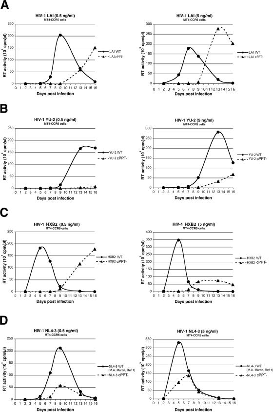
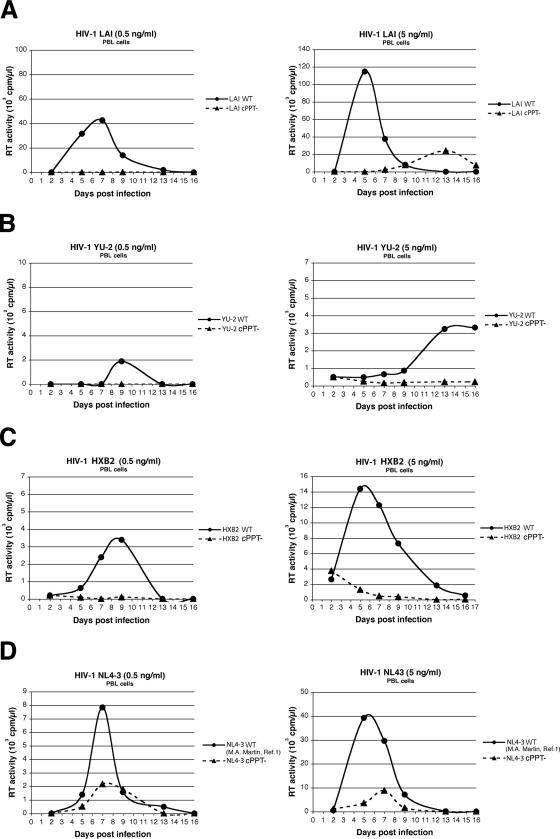
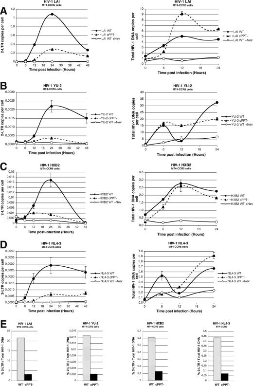
We have previously established, using human immunodeficiency virus type 1 (HIV-1) strain LAI, that the HIV-1 central DNA Flap acts as a cis determinant of viral genome nuclear import. Although the impact of the DNA Flap on nuclear import has already found numerous independent confirmations in the context of lentivirus vectors, it has been claimed that it may be nonessential for infectious virus strains LAI, YU-2 (J. D. Dvorin et al., J. Virol. 76:12087-12096, 2002), HXB2, and NL4-3 (A. Limon et al., J. Virol. 76:12078-12086, 2002). We conducted a detailed analysis of virus infectivity using the provirus clones provided by the authors and analogous target cells. In contrast to published data, our results show that all cPPT mutant viruses exhibit reduced infectivity corresponding to a nuclear import defect irrespective of the viral genetic background or target cell.


Figures

FIG. 1.
FIG. 1.
One-round titration assay of wild-type HIV-1 and cPPT mutants in dividing or nondividing P4-CCR5 indicator cells. (A to D) P4-CCR5 indicator cells were infected in triplicate with 20, 10, 5, or 2.5 ng of p24 antigen/ml (three times more for HXB2) from wild-type or cPPT mutant HIV-1 LAI (A), YU-2 (B), HXB2 (C), or NL4-3 (D). (E) Cell cycle arrest was induced by aphidicolin treatment (8 μM) from the day prior to infection, and nondividing P4-CCR5 cells were infected in triplicate with 20 ng of p24 antigen/ml from wild-type or cPPT mutant HIV-1 LAI, YU-2, HXB2, or NL4-3. β-Galactosidase activity was measured 48 h postinfection and was expressed as mean relative light units (RLU)/s (A to D) or RLU/s/ng of p24 (E) ± standard deviation. Data shown are representative of three independent experiments.
FIG. 2.
FIG. 2.
Replication kinetics of wild-type HIV-1 and cPPT mutants in MT4-CCR5 lymphocytes. MT4-CCR5 cells were infected with 0.5 or 5 ng of p24 antigen/ml (left and right panels, respectively) from wild-type or cPPT mutant HIV-1 LAI (A), YU-2 (B), HXB2 (C), or NL4-3 (D). Reverse transcriptase (RT) activity was measured on culture supernatants collected at regular time points over 16 days postinfection and is expressed in counts per minute per microliter. Results are representative of two independent experiments.
FIG. 3.
FIG. 3.
Replication kinetics of wild-type HIV-1 and cPPT mutants in PBLs. PBLs were infected with 0.5 or 5 ng of p24 antigen/ml (left and right panels, respectively) from wild-type or cPPT mutant HIV-1 LAI (A), YU-2 (B), HXB2 (C), or NL4-3 (D). Reverse transcriptase (RT) activity was measured on culture supernatants collected at regular time points over 16 days postinfection and is expressed in counts per minute per microliter. Results are representative of two independent experiments.
FIG. 4.
FIG. 4.
Quantitative PCR analysis of 2-LTR circle formation and total HIV-1 DNA copy number following infection of MT4-CCR5 cells with wild-type HIV-1 or cPPT mutants. (A to D) MT4-CCR5 cells were infected with wild-type or cPPT mutant LAI (A), YU-2 (B), HXB2 (C), or NL4-3 (D), and total DNA was extracted at 0, 6, 12, 24, and 48 h postinfection. Quantification of 2-LTR circles (A to D, left panels) and total HIV-1 DNA (A to D, right panels) was performed by real-time PCR at each time point. Cells infected with wild-type viruses in the presence of nevirapine (+Nev) served as controls. Results are means of triplicate (2-LTR) and duplicate (pol) determinations ± standard deviations. (E) Graphs show the ratio of the copy number of 2-LTR circles to that of total HIV-1 DNA for each virus. The ratio was calculated by dividing the peak of the 2-LTR copy number by the peak of total viral DNA.

Similar articles

Cited by

References

    1. Adachi, A., H. E. Gendelman, S. Koenig, T. Folks, R. Willey, A. Rabson, and M. A. Martin. 1986. Production of acquired immunodeficiency syndrome-associated retrovirus in human and nonhuman cells transfected with an infectious molecular clone. J. Virol. 59:284-291. - PMC - PubMed
    1. Ao, Z., X. Yao, and E. A. Cohen. 2004. Assessment of the role of the central DNA flap in human immunodeficiency virus type 1 replication by using a single-cycle replication system. J. Virol. 78:3170-3177. - PMC - PubMed
    1. Baekelandt, V., A. Claeys, K. Eggermont, E. Lauwers, B. De Strooper, B. Nuttin, and Z. Debyser. 2002. Characterization of lentiviral vector-mediated gene transfer in adult mouse brain. Hum. Gene Ther. 13:841-853. - PubMed
    1. Barry, S. C., B. Harder, M. Brzezinski, L. Y. Flint, J. Seppen, and W. R. Osborne. 2001. Lentivirus vectors encoding both central polypurine tract and posttranscriptional regulatory element provide enhanced transduction and transgene expression. Hum. Gene Ther. 12:1103-1108. - PubMed
    1. Bouyac-Bertoia, M., J. D. Dvorin, R. A. Fouchier, Y. Jenkins, B. E. Meyer, L. I. Wu, M. Emerman, and M. H. Malim. 2001. HIV-1 infection requires a functional integrase NLS. Mol. Cell 7:1025-1035. - PubMed

Publication types

LinkOut - more resources