. 2006 Sep 27;26(39):9902-12.
doi: 10.1523/JNEUROSCI.2897-06.2006.

Beta-arrestin2-mediated internalization of mammalian odorant receptors

Affiliations

Beta-arrestin2-mediated internalization of mammalian odorant receptors

Anastasia Mashukova et al. J Neurosci. .

Abstract

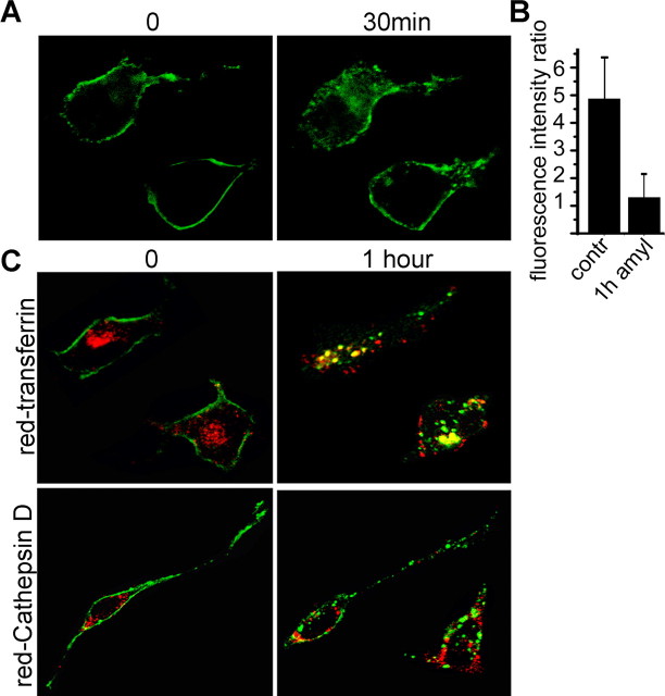
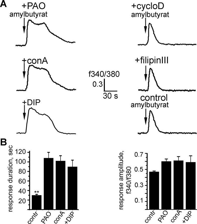
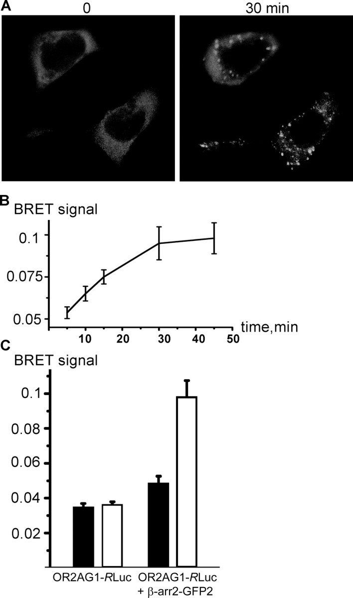
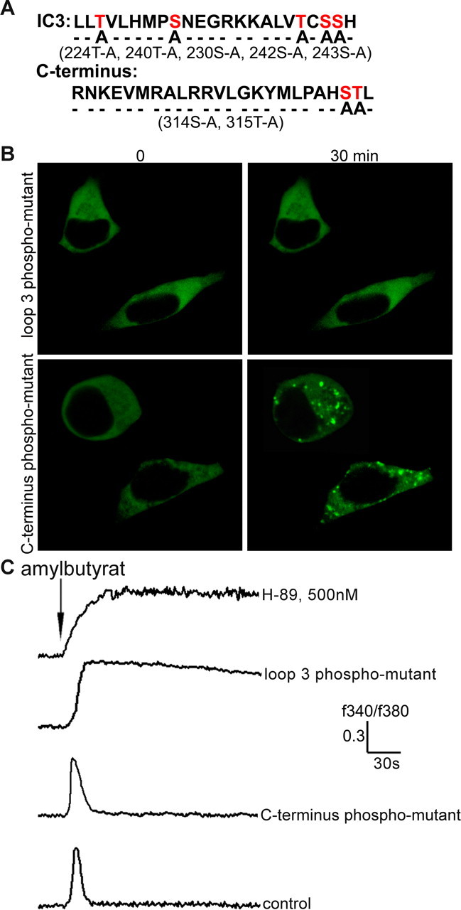
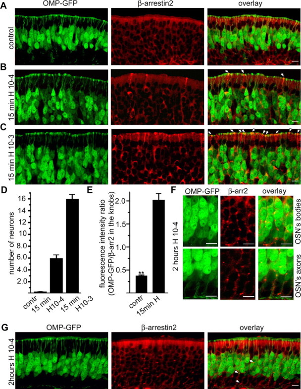
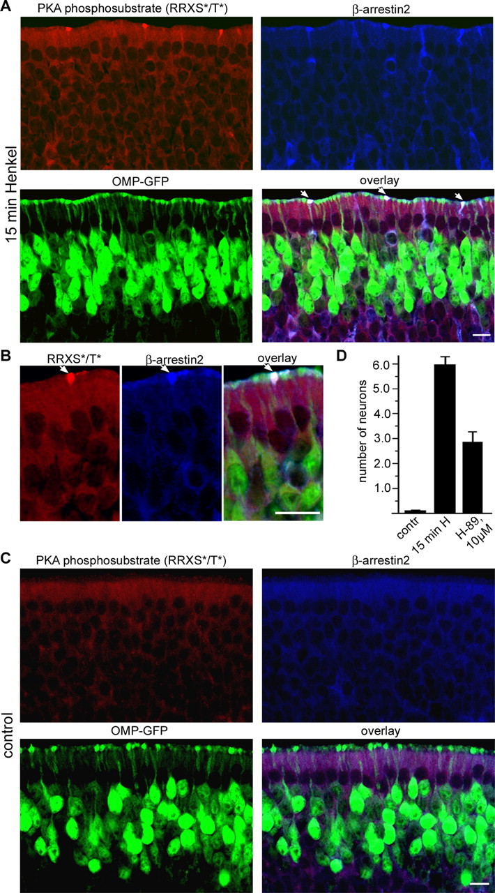
Odorant receptors comprise the biggest subfamily of G-protein-coupled receptors. Although the endocytic mechanisms of other G-protein-coupled receptors have been characterized extensively, almost nothing is known about the intracellular trafficking of odorant receptors. The present study describes the endocytic pathway of mammalian odorant receptors, which bind beta-arrestin2 with high affinity and are internalized via a clathrin-dependent mechanism. After prolonged odorant exposure, receptors are not targeted to lysosomal degradation but accumulate in recycling endosomes. Odorant-induced odorant receptor desensitization is promoted by cAMP-dependent protein kinase A phosphorylation and is dependent on serine and threonine residues within the third intracellular loop of the receptor. Moreover, beta-arrestin2 is redistributed into the dendritic knobs of mouse olfactory receptor neurons after treatment with a complex odorant mixture. Prolonged odorant exposure resulted in accumulation of beta-arrestin2 in intracellular vesicles. Adaptation of olfactory receptor neurons to odorants can be abolished by the inhibition of clathrin-mediated endocytosis, showing the physiological relevance of the here described mechanism of odorant receptor desensitization. A better understanding of odorant receptor trafficking and additional insight into the molecular determinants underlying the interactions of odorant receptors with beta-arrestin2 and other trafficking proteins will therefore be important to fully understand the mechanisms of adaptation and sensitization in the olfactory epithelium.


Figures

Figure 1.
Figure 1.
OR2AG1 undergoes endocytosis during stimulation. A, Confocal microscopy of cells transfected with OR2AG1–GFP before (0) and after (30 min) amylbutyrat treatment. OR2AG1–GFP is redistributed from the cell membrane to intracellular vesicles during stimulation. B, Quantification of the OR2AG1–GFP fluorescence intensity in the membrane relative to the intensity in the cytosol, measured in single confocal sections before (contr) and after the amylbutyrat (amyl) application. The averages of at least 15 cells demonstrate a ∼4.5 fold decrease in membrane GFP fluorescence intensity after stimulation. C, Confocal microscopy of HEK293 cells expressing OR2AG1–GFP before (0) and after (1 h) amylbutyrat stimulation. OR2AG1–GFP-containing vesicles (green) show substantial colocalization with transferrin (red, top). After 1 h of amylbutyrat stimulation (yellow, top), only marginal colocalization was observed with the lysosomal protease cathepsin D (red, bottom).
Figure 2.
Figure 2.
Endocytosis of OR2AG1 is mediated by a clathrin-dependent mechanism. A, Representative ratiofluorometric recordings of the individual cells. The cytosolic Ca2+ level of fura-2-loaded cells is depicted as fluorescence ratio (F340/F380) and viewed as a function of time. Cells were preincubated with inhibitors of the clathrin pathway [phenylarsine oxide (+PAO), concanavalinA (+conA), and dynamin inhibitory peptide (+DIP)] or the caveolae pathway [filipin III (+filipin III) and β-cyclodextrin (+cycloD)] as described in Materials and Methods. B, Preincubation of the cells with clathrin inhibitors resulted in significantly prolonged responses compared with the control, although response amplitude was only slightly increased. The duration of the Ca2+ response was measured from the onset of the stimulus to the decrease to the basal level. Results are the mean from at least 10 cells. Error bars represent SEM; **p < 0.01. Amylbutyrat (100 μm) was applied for 5 s in each case.
Figure 3.
Figure 3.
β-arrestin2 interacts with olfactory receptors during ligand binding. A, Confocal microscopy of HEK293 cells cotransfected with OR2AG1 and β-arrestin2–GFP. Amylbutyrat treatment (30 min) causes translocation of β-arrestin2 into intracellular vesicular structures. B, Cells were cotransfected with β-arrestin–GFP and OR2AG1–RLuc fusion constructs, and the BRET ratio was measured at different time points of amylbutyrat stimulation 48 h after transfection. The BRET signal increased in a time-dependent manner during addition of the agonist. Data are the mean from five independent experiments. C, Cells were cotransfected with β-arrestin–GFP and OR2AG1–RLuc fusion constructs or with OR2AG1–RLuc alone. The BRET ratio was determined 48 h after transfection on amylbutyrat-stimulated (white bars) and nonstimulated (black bars) cells. No significant change of the BRET ratio during addition of amylbutyrat is observed in cells expressing OR2AG1–RLuc alone. In contrast, addition of the agonist to cells expressing the OR2AG1–RLuc fusion construct together with β-arrestin–GFP leads to an increase of the BRET signal. Error bars represent SEM.
Figure 4.
Figure 4.
Agonist promoted OR phosphorylation is essential for OR desensitization and β-arrestin2 binding. A, Site-directed mutagenesis experiments were performed to delete the potential phosphorylation sites of OR2AG1; the scheme shows mutated phosphorylation sites within the third intracellular loop (positions 224T-A, 240T-A, 230S-A, 242S-A, and 243S-A) and the C terminus (positions 314S-A and 315T-A). B, OR2AG1 with mutated intracellular loop 3 failed to induce β-arrestin2–GFP translocation, whereas OR2AG1 with mutated C terminus shows wild-type-like β-arrestin2–GFP translocation into endosomes in response to amylbutyrat stimulation. C, Ca2+ responses elicited by the activation of the C-terminus phosphorylation mutant (C terminus phospho-mutant) in HEK293 cells were undistinguishable from the control. In contrast, a strong delay in desensitization was found for the third loop phosphorylation mutant (loop 3 phospho-mutant). OR2AG1 desensitization is promoted by PKA phosphorylation, as reflected by a strong delay in desensitization in the presence of the PKA inhibitor H-89 (500 nm). These delays in desensitization were observed in at east 10 independent experiments. Amylbutyrat (100 μm) was applied for 5 s in each case.
Figure 5.
Figure 5.
β-Arrestin2 is redistributed in ORNs during odorant treatment. A–C, β-Arrestin2 redistribution in mouse ORNs after odorant exposure. Fifteen-day-old OMP–GFP mice were exposed or not (control) to a complex odorant mixture of 100 compounds (Henkel 100) for 15 min. Cryosections through the mouse olfactory epithelium were probed with antibodies against β-arrestin2. A, In untreated mice (control), β-arrestin2 was homogeneously distributed throughout the cytoplasm of all cells in the olfactory epithelium, including ORNs (yellow on the overlay). B, Fifteen minutes of exposure to the Henkel 100 mixture (diluted 1:10,000) caused translocation of β-arrestin2 to the dendritic knobs of ORNs as indicated by arrows. C, Treatment of mice with higher concentration of Henkel 100 mixture (1:1000) resulted in increased number of ORNs with β-arrestin2 accumulation in the dendritic knob. D, E, Quantification of the observed effects; three mice from the control and three mice from the exposed group were investigated (4 slices with complete olfactory epithelium per mouse). D, The number of neurons showing strong anti-β-arrestin2 reactivity in the dendritic knob was counted over a 500 μm segment of a 10-μm-thick cryosection from the olfactory epithelium. Error bars represent SEM. E, Quantification of the anti-β-arrestin2 fluorescence intensity in the dendritic knobs of ORNs relative to the fluorescence intensity from OMP–GFP. The comparison of the fluorescence intensities in the dendritic knobs from the ORNs from the control group and the intensities from the group exposed to the lower Henkel 100 concentration demonstrates an approximate sixfold increase. At least 70 neurons from each group were investigated. Error bars represent SD; **p < 0.01. F, Higher-magnification pictures show a redistribution of β-arrestin2 to vesicular structures in the cell bodies (OSN‘s bodies) and in the axons (OSN‘s axons) of ORNs after 2 h odorant treatment. G, After 2 h of odorant treatment (Henkel 100 mixture, 2 h Henkel), β-arrestin2 showed vesicular-like staining in the ORNs as indicated by arrows. Scale bars, 10 μm.
Figure 6.
Figure 6.
PKA is involved in β-arrestin2 accumulation in the dendritic knobs. A, Fifteen-day-old OMP–GFP mice were exposed to a complex odorant mixture of 100 compounds (Henkel 100) for 15 min, and cryosections through the olfactory epithelium were costained with antibodies against β-arrestin2 (blue) and antibodies that detect PKA phosphorylated proteins, motif RRXS*/T* (red). Strong anti-PKA phosphosubstrate immunoreactivity was observed in the cytoplasm and dendritic knobs of ORNs after odorant treatment (top left). In addition, ∼70% of the PKA phosphosubstrate-positive knobs also show strong anti-β-arrestin2 immunoreactivity (indicated by arrows on the overlay). B, Higher-magnification pictures show colocalization of PKA phosphorylated proteins (RRXS*/T*) and β-arrestin2 in the dendritic knobs of ORNs after 15 min of Henkel 100 treatment. Scale bars, 10 μm. C, Without odorant application, β-arrestin2 accumulation in the dendritic knobs and staining with the anti-PKA phosphosubstrate was not observed. D, Pretreatment of olfactory epithelium with 10 μm of the PKA inhibitor H-89 decreases the number of β-arrestin2-positive dendritic knobs after 15 min of Henkel 100 mixture treatment. Septal bone with the intact olfactory epithelium was dissected from the head and preincubated for 15 min in the large volume of Ringer’s solution (control, 15 min H) or in Ringer’s solution containing 10 μm of the PKA inhibitor H-89 (10 μm). After that, the olfactory epithelium was placed in 1:10,000 diluted Henkel 100 mixture (15 min H; H-89, 10 μm) or in Ringer’s solution (control) for 15 min and fixed. Cryosections from olfactory epithelium were stained with anti-β-arrestin2 antibodies. The number of neurons showing strong anti-β-arrestin2 reactivity in the dendritic knob was counted per 500 μm of olfactory epithelium surface. Error bars represent SEM. Scale bars, 10 μm.
Figure 7.
Figure 7.
Endocytosis is important for adaptation of ORNs. A, A 2 h pretreatment of mice with Henkel 100 mixture results in significantly reduced amplitude of EOG responses. Twelve mice were pretreated with Henkel 100 mixture for 2 h, and 11 mice did not receive any treatment (control group). The septal bone with the intact olfactory epithelium was dissected from the head and used intact for EOG recording as described in Materials and Methods. Increasing concentrations of Henkel 100 mixture were applied in the vapor phase for 2 s, with 1 min interval. Bars are the mean amplitude of the EOG responses from the control group (shown in white) and Henkel 100 pretreated mice (shown in black). Factorial ANOVA was performed. Error bars represent SEM; *p < 0.001. B, Treatment of mouse olfactory epithelium with Henkel 100 mixture enhances uptake of fluid phase marker HRP. Olfactory epithelium was dissected from the septal bone and incubated in solution containing 0.25 mg/ml HRP in Ringer’s solution (control; squares) or in Ringer’s solution containing Henkel 100 mixture at 1:10,000 dilution (circles) for indicated periods of time at room temperature. After the incubation, the olfactory epithelium was washed and lysed, and HRP activity was determined as described in Material and Methods. Each point is the mean from three independent experiments. Error bars represent SEM. C, D, Inhibition of clathrin-mediated endocytic pathway affects sensory adaptation in ORNs. C, A representative ratiofluorometric recording of the cytosolic Ca2+ level (fluorescence ratio, F340/F380, viewed as a function of time) shows the effect of dynamin inhibitory peptide (5 min incubation; DIP), resulting in a significant loss of adaptation. Arrows indicate repeated stimulation with a complex odorant mixture (Henkel 50 B, 5 s each) at changing intervals. D, Fluorescence image of an acutely dissociated ORN loaded with the ratiometric Ca2+ dye fura-2. Neurons are easily identified based on their characteristic morphology. Changes in Ca2+ concentration were measured in the dendritic knob region (depicted by the circle). E, Quantification of the observed effects. Bars represent the average residual response normalized to an initial odorant application as a function of the prestimulation interval. Overall, 17 individual ORNs were included in the analysis. Under control conditions, all of these neurons responded with decreasing signal amplitudes to repeated stimulation. At interstimulus intervals, ≤40 s residual response amplitudes were 57.65 ± 2.14% (control; n = 10) and 78.48 ± 8.85% (treatment; n = 6), whereas at intervals, ≤60 s residual amplitudes were 81.77 ± 4.63% (control; n = 5) and 104.13 ± 5.51% (treatment; n = 5). Response amplitudes before and after pharmacological treatment were statistically analyzed using a homoscedastic Student’s t test. Error bars represent SEM; * p < 0.1; **p < 0.01.

Similar articles

Cited by

References

    1. Arshavsky VY. Protein translocation in photoreceptor light adaptation: a common theme in vertebrate and invertebrate vision. Sci STKE. 2003;2003:E43. - PubMed
    1. Bannister LH, Dodson HC. Endocytic pathways in the olfactory and vomeronasal epithelia of the mouse: ultrastructure and uptake of tracers. Microsc Res Tech. 1992;23:128–141. - PubMed
    1. Barnea G, O’Donnell S, Mancia F, Sun X, Nemes A, Mendelsohn M, Axel R. Odorant receptors on axon termini in the brain. Science. 2004;304:1468. - PubMed
    1. Bhandawat V, Reisert J, Yau KW. Elementary response of olfactory receptor neurons to odorants. Science. 2005;308:1931–1934. - PMC - PubMed
    1. Boekhoff I, Schleicher S, Strotmann J, Breer H. Odor-induced phosphorylation of olfactory cilia proteins. Proc Natl Acad Sci USA. 1992;89:11983–11987. - PMC - PubMed

Publication types

MeSH terms

LinkOut - more resources