. 2006 Sep 27;26(39):9944-55.
doi: 10.1523/JNEUROSCI.2990-06.2006.

Vestibular hair bundles control pH with (Na+, K+)/H+ exchangers NHE6 and NHE9

Affiliations

Vestibular hair bundles control pH with (Na+, K+)/H+ exchangers NHE6 and NHE9

Jennifer K Hill et al. J Neurosci. .

Abstract

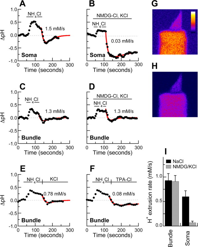
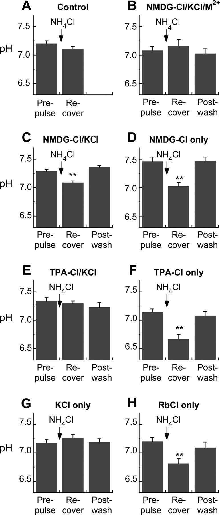
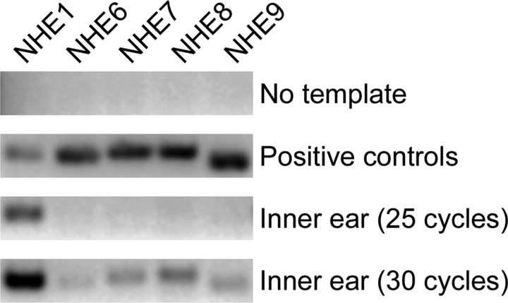
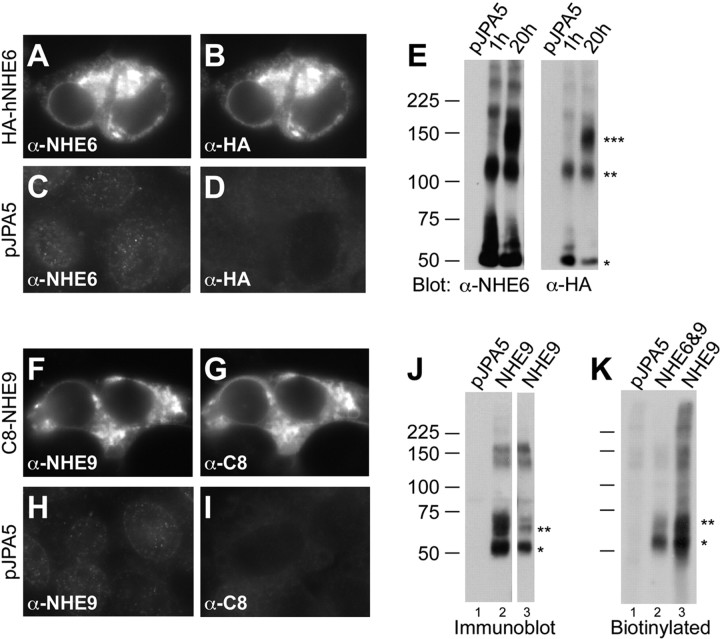
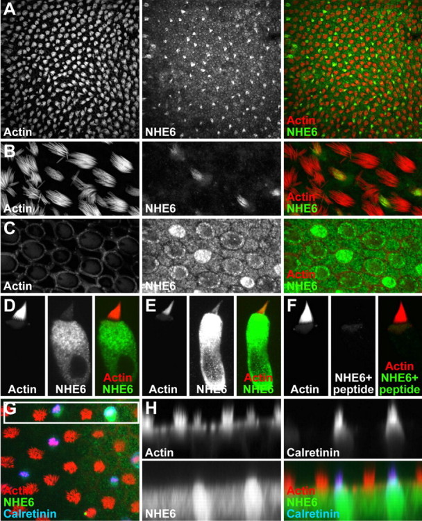
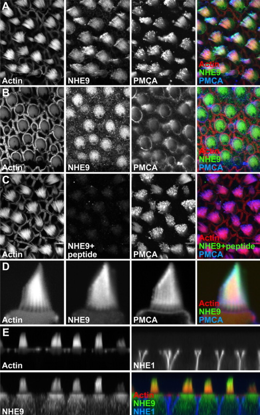
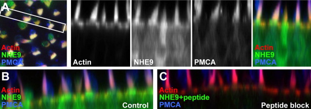
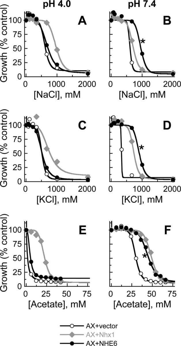
In hair cells of the inner ear, robust Ca2+/H+ exchange mediated by plasma-membrane Ca2+-ATPase would rapidly acidify mechanically sensitive hair bundles without efficient removal of H+. We found that, whereas the basolateral membrane of vestibular hair cells from the frog saccule extrudes H+ via an Na+-dependent mechanism, bundles rapidly remove H+ in the absence of Na+ and HCO3(-), even when the soma is acidified. K+ was fully effective and sufficient for H+ removal; in contrast, Rb+ failed to support pH recovery. Na+/H+-exchanger isoform 1 (NHE1) was present on hair-cell soma membranes and was likely responsible for Na+-dependent H+ extrusion. NHE6 and NHE9 are organellar isoforms that can appear transiently on plasma membranes and have been proposed to mediate K+/H+ exchange. We identified NHE6 in a subset of hair bundles; NHE9 was present in all bundles. Heterologous expression of these isoforms in yeast strains lacking endogenous exchangers conferred pH-dependent tolerance to high levels of KCl and NaCl. NHE9 preferred cations in the order K+, Na+ >> Rb+, consistent with the relative efficacies of these ions in promoting pH recovery in hair bundles. Electroneutral K+/H+ exchange, which we propose is performed by NHE9 in hair bundles, exploits the high-K+ endolymph, responds only to pH imbalance across the bundle membrane, is unaffected by the +80 mV endocochlear potential, and uses mechanisms already present in the ear for K+ recycling. This mechanism allows the hair cell to remove H+ generated by Ca2+ pumping without ATP hydrolysis in the cell.


Figures

Figure 1.
Figure 1.
Dynamic pH measurements in bundles and somas of isolated hair cells. Change in pH from resting pH is plotted. A, B, Soma pH in response to NH4Cl/NaCl pulse and washes with standard saline (A) or NMDG-Cl, 2 mm KCl, and divalent cations (B). C–F, Hair bundle pH in response to NH4Cl/NaCl pulse and washes with standard saline (C), NMDG-Cl/KCl/M2+ (D), KCl only (E), or TPA-Cl only (F). G, H, Protonated (G) and unprotonated (H) SNARF-5 signal in resting hair cell. Because of the low volume in the confocal section, the bundle signal is low compared with that in the soma. The linear color scale indicated in G also applies to H. I, Recovery rates of bundle and soma pH after acidification.
Figure 2.
Figure 2.
Static pH measurements in hair bundles of isolated hair cells. For each condition, pH was measured before (Prepulse) and 2–3 min after (Recover) a 25 mm NH4Cl pulse; pH was subsequently measured after a wash with standard saline (Post-wash). **p < 0.001, significance between Prepulse and Recover. Results displayed are mean ± SE from at least two dishes of isolated cells. A, Standard saline recovery (no Post-wash). B, NMDG-Cl/KCl/M2+. C, NMDG-Cl/KCl. D, NMDG-Cl only. E, TPA-Cl/KCl. F, TPA-Cl only. G, KCl only. H, RbCl only.
Figure 3.
Figure 3.
RT-PCR detection of NHE isoforms in mouse inner ear. NHE isoforms are indicated on top; template source is indicated on right. Positive controls were plasmids with appropriate NHE fragment. Masses of products were as expected (NHE1, 149 bp; NHE6, 145 bp; NHE7, 153 bp; NHE8, 158 bp; NHE9, 140 bp).
Figure 4.
Figure 4.
Specificity of NHE6 and NHE9 antibodies. A–D, Immunocytochemistry with anti-NHE6. Cell expressing HA-tagged human NHE6 (A, B) or control plasmid (C, D) were detected with anti-NHE6 (A, C) or anti-HA tag (B, D) antibody. E, Immunoblot with anti-NHE6. COS-7 cells expressing control plasmid (left lanes in each blot) or HA-tagged human NHE6 (right two lanes) were probed with anti-NHE6 (left blot) or anti-HA (right blot). Increased incubation of the cell extract with SDS sample buffer led to an increase in a high-molecular-weight band (***) at 20 h compared with 1 h. At shorter times, bands likely corresponding to monomer (*) and dimer (**) are most prominent. F–I, Immunocytochemistry with anti-NHE9. Cell expressing C8-tagged mouse NHE9 (F, G) or control plasmid (H, I) were detected with anti-NHE9 (F, H) or anti-C8 tag (G, I). J, Immunoprecipitation with anti-NHE9 (lanes 1, 2) or with anti-C8 (lane 3), followed by immunoblotting with anti-C8 (lanes 1, 2) or anti-NHE9 (lane 3). K, Detection of plasma-membrane NHE9 by surface biotinylation. COS-7 cells transfected with indicated plasmids were biotinylated with a membrane-impermeable biotinylation reagent; after immunoprecipitation with anti-NHE9, proteins were detected with avidin–alkaline phosphatase.
Figure 5.
Figure 5.
Localization of NHE6 in bullfrog saccule. A, Low-power view of frog saccule labeled with phalloidin (for actin) and anti-NHE6. Only a small fraction of cells are positive. B, High-power view of frog saccule. Small bundles are detected with anti-NHE6. C, Same field as B but at the level of the cuticular plate. Cells with high levels of NHE6 in their bundles have more NHE6 in the soma. D, Isolated hair cell with large hair bundle. Little NHE6 is present in the bundle. E, Isolated hair cell with narrow hair bundle. NHE6 labeling is strong in the soma and modest in the bundle. F, Isolated hair cell labeled after preincubation of anti-NHE6 antibody with antigenic peptide. NHE6 labeling is abolished. G, Single confocal section of z-series triple labeled with phalloidin, anti-NHE6, and anti-calretinin. Box indicates region used for x–z reslice. H, x–z reslice from z-series represented by G. Note that cells labeled strongly with NHE6 are also positive for calretinin. Full-frame widths: A, 183 μm; B, C, 59 μm; D–F, 18 μm; G, H, 62 μm.
Figure 6.
Figure 6.
Localization of NHE9 in bullfrog saccule. A, Low-power view of frog saccule, at level of hair bundles, labeled with phalloidin (for actin), anti-NHE9, and 5F10 (for all PMCA isoforms). All bundles have NHE9 immunoreactivity. B, Same field as A but at the level of the cuticular plate. Hair cells (but not supporting cells) have substantial NHE9 immunoreactivity. C, Frog saccule labeled after preincubation of anti-NHE9 antibody with antigenic peptide. NHE9 labeling is abolished. D, High-power view of hair bundle from isolated frog hair cell. NHE9 labeling is throughout the bundle, with some concentration at tips of the stereocilia. E, x–z reslice of z-stack of frog saccule labeled with phalloidin, anti-NHE9, and anti-NHE1. Note that NHE1 is found on hair-cell basolateral membranes. Full-frame widths: A–C, 46 μm; D, 10 μm; E, 65 μm.
Figure 7.
Figure 7.
Localization of NHE9 in rat utricle. A, Left, Single confocal section of rat utricle z-series triple labeled with phalloidin, anti-NHE9, and 5F10 (for PMCA). The box indicates the region used for x–z reslice. Right, Individual channels and merged image from x–z reslice. B, C, x–z reslices of rat utricles labeled with phalloidin, anti-NHE9, and 5F10 either without (B) or with (C) the NHE9 antigenic peptide. Full-panel widths: A, 36 μm; B, C, 78 μm.
Figure 8.
Figure 8.
NHE6 expression reduces sensitivity of yeast growth to salt and weak acid stress. AX strain (lacking NHA1 and NHX1 ion transporters) was transformed with empty vector, yeast Nhx1, or human NHE6. Growth was assessed in the presence of varying concentrations of the following: A, NaCl at pH 4.0; B, NaCl at pH 7.4; C, KCl at pH 4.0; D, KCl at pH 7.4; E, sodium acetate at pH 4.0; and F, sodium acetate at pH 7.4. NHE6 rescued the slow-growth phenotype at pH 7.4 (*). Data points are averages of eight replicates combined from two independent experiments. Nearly identical results were obtained with two independently transformed clones of NHE6.
Figure 9.
Figure 9.
NHE9 expression reduces sensitivity of yeast growth to salt. AX strain was transformed with empty vector, yeast Nhx1, mutant Y361F Nhx1, or mouse NHE9. Growth of wild type yeast (WT), AX, or transformants was assessed in the presence of varying concentrations of the following: A, NaCl at pH 4.0; B, NaCl at pH 7.4; C, KCl at pH 4.0; D, KCl at pH 7.4; E, LiCl at pH 4.0; F, LiCl at pH 7.4; E, RbCl at pH 4.0; and F, RbCl at pH 7.4. NHE9 rescued the slow-growth phenotype at pH 7.4 (*). Data points are averages of two to four replicates.

References

    1. Abacioglu YH, Fouts TR, Laman JD, Claassen E, Pincus SH, Moore JP, Roby CA, Kamin-Lewis R, Lewis GK. Epitope mapping and topology of baculovirus-expressed HIV-1 gp160 determined with a panel of murine monoclonal antibodies. AIDS Res Hum Retroviruses. 1994;10:371–381. - PubMed
    1. Aronson PS. Kinetic properties of the plasma membrane Na+-H+ exchanger. Annu Rev Physiol. 1985;47:545–560. - PubMed
    1. Blair HC, Teitelbaum SL, Ghiselli R, Gluck S. Osteoclastic bone resorption by a polarized vacuolar proton pump. Science. 1989;245:855–857. - PubMed
    1. Bond BR, Ng LL, Schulte BA. Identification of mRNA transcripts and immunohistochemical localization of Na/H exchanger isoforms in gerbil inner ear. Hear Res. 1998;123:1–9. - PubMed
    1. Bowman EJ, Siebers A, Altendorf K. Bafilomycins: a class of inhibitors of membrane ATPases from microorganisms, animal cells, and plant cells. Proc Natl Acad Sci USA. 1988;85:7972–7976. - PMC - PubMed

Publication types

MeSH terms