. 2006 Sep 27;26(39):9975-82.
doi: 10.1523/JNEUROSCI.2595-06.2006.

Targeted deletion of a single Sca8 ataxia locus allele in mice causes abnormal gait, progressive loss of motor coordination, and Purkinje cell dendritic deficits

Affiliations

Targeted deletion of a single Sca8 ataxia locus allele in mice causes abnormal gait, progressive loss of motor coordination, and Purkinje cell dendritic deficits

Yungui He et al. J Neurosci. .

Abstract

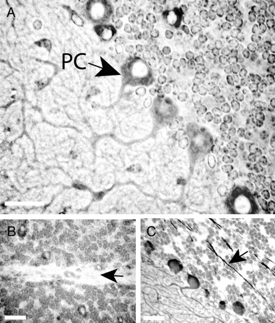
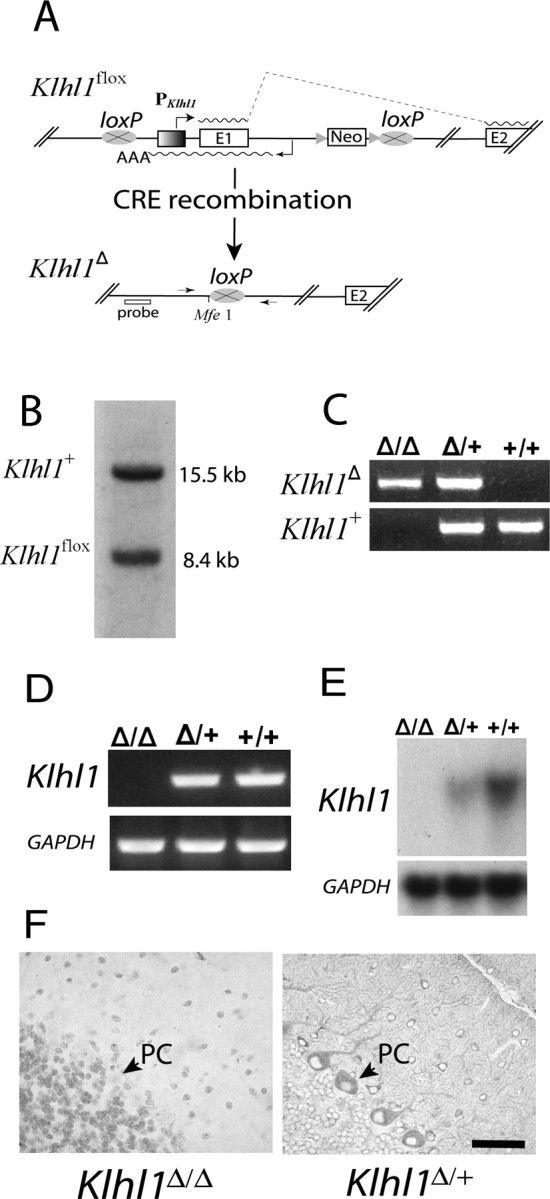
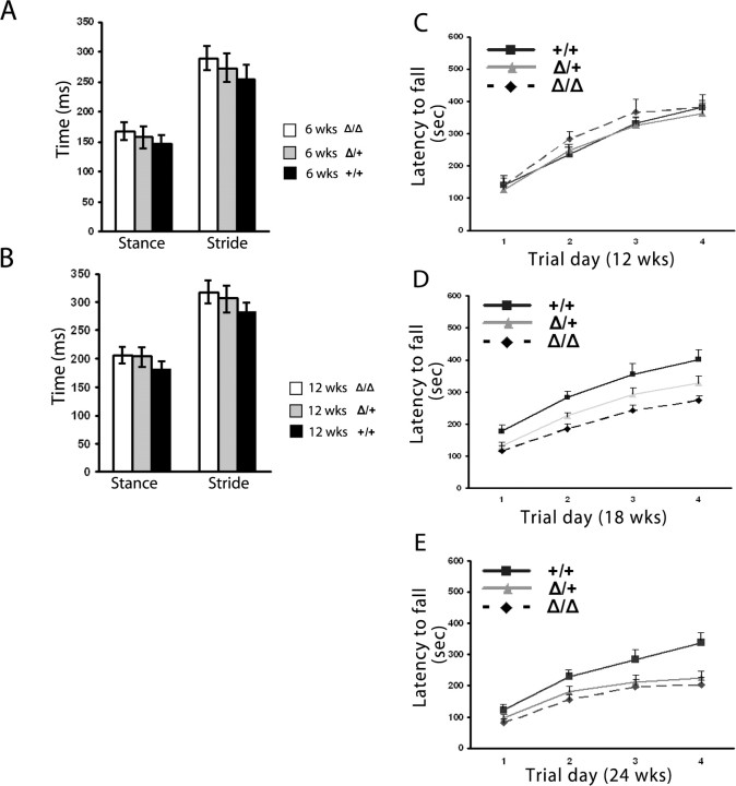
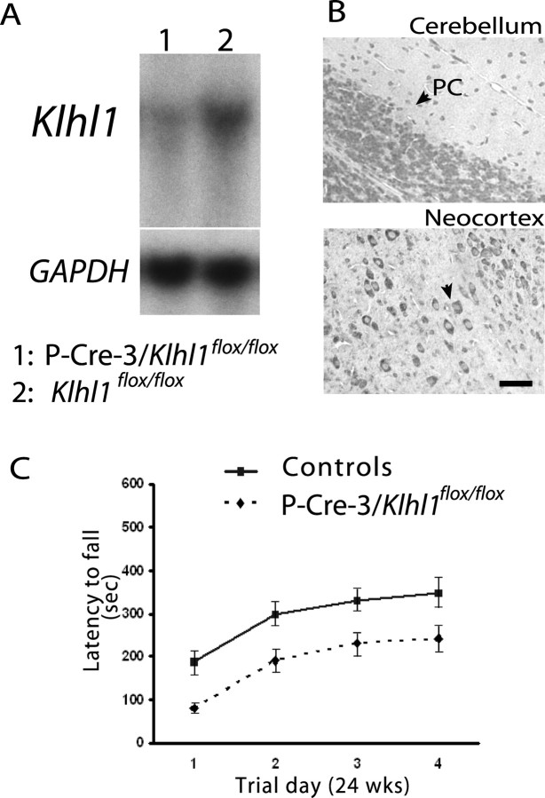
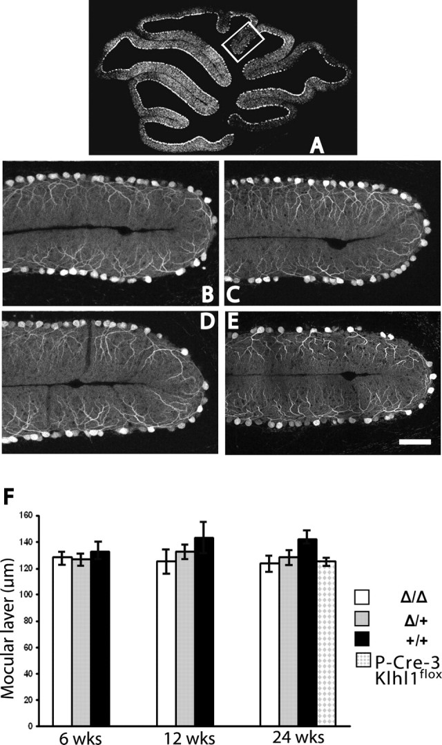
Spinocerebellar ataxia type 8 (SCA8) patients typically have a slowly progressive, adult-onset ataxia. SCA8 is dominantly inherited and is caused by large CTG repeat expansions in the untranslated antisense RNA of the Kelch-like 1 gene (KLHL1), but the molecular mechanism through which this expansion leads to disease is still unknown. To more fully characterize the underlying molecular mechanisms involved in SCA8, we developed a mouse model in which Klhl1 is deleted in either all tissues or is deleted specifically in Purkinje cells only. We found that mice that are either homozygous or heterozygous for the Klhl1 deletion have significant gait abnormalities at an early age and develop a significant loss of motor coordination by 24 weeks of age. This loss progresses more rapidly in homozygous knock-outs. Mice with Klhl1 specifically deleted in only Purkinje cells had a loss of motor coordination that was almost identical to the total-tissue deletion mice. Finally, we found significant Purkinje cell dendritic deficits, as measured by the thickness of the molecular layer, in all mice in which Klhl1 was deleted (both total and Purkinje cell-specific deletions) and an intermediate reduction in molecular layer thickness in mice with reduced levels of Klhl1 expression (heterozygous deletions). The results from this mouse model show that even a partial loss of Klhl1 function leads to degeneration of Purkinje cell function and indicates that loss of KLHL1 activity is likely to play a significant part in the underlying pathophysiology of SCA8.


Figures

Figure 1.
Figure 1.
Cellular and subcellular localization of Klhl1 in mouse cerebellar tissue. Immunohistochemical staining of normal FVB mice at 4 weeks of age with KLHL1-specific monoclonal antibody 13D9 shows that Klhl1 is a prominent cytoplasmic protein in the soma and dendrites of cerebellar Purkinje cells but is not present in the nuclei (A) or the axons (B) of these cells. Both the axons and the nuclei of Purkinje cells stain positively with anti-calbindin antibody (C). PC, Purkinje cell. Black arrows show the axons of the Purkinje cells. Scale bars, 25 μm.
Figure 2.
Figure 2.
Expression of Klhl1 is eliminated in Klhl1Δ/Δ mice and is significantly reduced in Klhl1Δ/+ mice. A, Overview of the deletion strategy and of the modified loci Klhl1flox (floxed) and Klhl1Δ (deleted). Klhl1flox mice were generated by flanking the Klhl1 transcription start site and the first Klhl1 coding exon and Klhl1as gene with two loxP sites. The neomycin-resistant selection cassette (Neo) used in targeting was inserted into intron 1 of Klhl1 and flanked with two FRT sites, which were used to delete the cassette by mating the mice to FLP transgenic animals. The Klhl1flox allele was then modified by mating to cre transgenic mice to generate mouse lines carrying a Klhl1 null allele (Klhl1Δ) in either all tissues (ubiquitous CRE expression) or specific tissues only (tissue-specific CRE expression). E, Exon; PKlhl1, Klhl1 promoter; triangle, FRT site; oval with a cross, loxP recombination sites; waved line, mRNA, AAA, polyadenylation signal of Klhl1as. The arrows in the bottom indicate the two primers that cross the 5′ and 3′ loxP sites (MEX1–8R and MEX2–3, indicated by P1 and P2), and the approximate location of the probe by Southern blot analysis (below) is also shown. B, Southern blot analysis of a heterozygous mouse showing both the wild-type (Klhl1+) and the Klhl1flox allele. The Southern blot was performed on MfeI-digested tail DNA using a flanking probe generated the expected 8.4 and 15.5 kb fragments. C, The genotyping of Klhl1Δ/Δ, Klhl1Δ/+, and Klhl1+/+ mice was performed by PCR using mouse tail DNA. Primers MEX1–8R and MEX2–3 generate a 642 bp fragment, whereas primer set MEXKO5C and MEX1–8R generate a 557 bp product from the wild-type Klhl1+ allele. D, RT-PCR analysis. RNA isolated from cerebella of 3-week-old mice was reverse transcribed and assayed by PCR using primer pair Mus2–22 and Mus3–24R. The mRNA of Klhl1 is clearly absent in the homozygous Klhl1Δ/Δ mice (lane 1), whereas in the heterozygous Klhl1Δ/+ (lane 2) and wild-type Klhl1+/+ (lane 3) cerebella, the primers are able to detect a 682 bp amplification product representing the Klhl1 cDNA. E, Northern blot analysis of Klhl1 mRNA in adult knock-out mice and their wild-type controls. Messenger RNA of Klhl1 is clearly absent in Klhl1Δ/Δ mice, and the expression is dramatically decreased in the Klhl1Δ/+ mice compared with that of the wild-type Klhl1+/+ controls. A subsequent probe of GAPDH mRNA was used as a loading control. F, Paraffin cerebella sections from 3-week-old Klhl1Δ/Δ (left) and Klhl1Δ/+ (right) mice stained for Klhl1 using 13D9 mAb. Sections show loss of immunoreactivity in the Purkinje cells of homozygous Klhl1Δ/Δ mice (left), but Klhl1-positive staining is clearly visible in the cell soma and dendrites of Purkinje cells from the heterozygous Klhl1Δ/+ mice (right). Note that there is no hematoxylin counterstaining in the right. PC, Purkinje cell. Scale bars, 50 μm.
Figure 3.
Figure 3.
Gait analysis with a TreadScan device and performance on an accelerating rotarod detects progressive deficiencies in heterozygous Klhl1Δ/+ and homozygous Klhl1Δ/Δ mice. Two groups of mice were trained for 2 weeks, and the gait analysis was performed at a speed of 20 cm/s for 20 s. At 6 weeks of age (A), the stride time for heterozygous Klhl1Δ/+ and homozygous Klhl1Δ/Δ mice was significantly longer than wild-type siblings in both front paws and rear paws. At the age of 12 weeks (B), the abnormalities of gait is also significant for both front paws and rear paws. For stance time, 6-week-old homozygous mice have a significantly longer stance time than wild-type animals, whereas the heterozygous mice have an obvious but not yet quite significantly longer stance than the controls. For the 12-week-old group, gait abnormalities are more obvious; the homozygous and heterozygous mice have increased significant impairments of their stance time compared with wild-type controls. The gait results for the hindpaw only are shown in the graphs in this figure. Twelve-week-old (C), 18-week-old (D), and 24-week-old (E) mice were tested for four trials per day for 4 consecutive days on an accelerating rotarod. At 12 weeks of age, the performances of the homozygous Klhl1Δ/Δ and heterozygous Klhl1Δ/+ mice were indistinguishable with their wild-type controls, but, at 18 weeks of age, homozygous Klhl1Δ/Δ mice showed significantly impaired performance improvement (p = 0.002) whereas the heterozygous Klhl1Δ/+ mice showed obvious but not significant impairment (p = 0.052). At 24 weeks of age, both homozygous and heterozygous mice showed significant impaired performance improvements at day 4 (p < 0.001; p < 0.05 respectively). Error bars indicate SEM.
Figure 4.
Figure 4.
Analyses of mice in which Klhl1 is deleted only in cerebellar Purkinje cells. A, Northern blot analysis demonstrated that the mRNA Klhl1 is dramatically decreased in cerebellar tissue from 24-week-old homozygous P-CRE-3/Klhl1flox/flox mice (lane 1) compared with the controls (Klhl1flox/flox; lane 2). The RNA loading levels of each lane were measured by a subsequent hybridization with GAPDH probe. B, Paraffin-embedded sections of brain from homozygous P-CRE-3/Klhl1flox/flox mice stained for Klhl1 using 13D9 mAb show cerebellar Purkinje cell-specific Klhl1 deficits. There was an absence of staining of Klhl1 in the soma and dendrites of Purkinje cells (left), but Klhl1 is normally expressed in the other neurons in this brain (neocortex; right). PC, Purkinje cell. Scale bar, 25 μm. C, Motor coordination performance deficits of 24-week-old P-CRE-3/Klhl1flox/flox mice on accelerating rotarods. Their performance was significantly worse (p = 0.011 at day 4) than the performance of controls. Error bars indicate SEM.
Figure 5.
Figure 5.
Klhl1 knock-out mice have significant thinning of the cerebellar molecular layer. A–E, Immunohistochemical analysis of cerebellar sections of Klhl1 knock-out mice. A, Diagram of a midsagittal cerebellar section indicating the posterior location of the folia presented in B–E. B–E, Calbindin immunoassays of 24-week-old mice were used measure the thickness of cerebellar molecular layers of Klhl1Δ/Δ (B), Klhl1Δ/+ (C), and P-CRE-3/Klhl1flox/flox (E) mice were significantly thinner than in the wild-type controls (D). Scale bar: B–E, 50 μm. F, Relative thickness of the cerebellar molecular layer in Klhl1 knock-out mice at 6, 12, and 24 weeks of age. Both homozygous and heterozygous show no significant difference when they are at the age of 6 or 12 weeks old compared with the wild-type controls. However, at the age of 24 weeks, the thickness of the molecular layer of homozygous Klhl1Δ/Δ and heterozygous Klhl1Δ/+mice were significantly decreased (mean ± SD, 123.5 ± 11.9, p = 0.015, n = 6; 128.4 ± 10.6, p = 0.048, n = 6; and 125.1 ± 1.8, p = 0.035, n = 2) compared with the wild-type Klhl1+/+controls (142.2 ± 7.3; n = 3). Error bars in F indicate SEM.

Similar articles

Cited by

References

    1. Albagli O, Dhordain P, Deweindt C, Lecocq G, Leprince D. The BTB/POZ domain: a new protein-protein interaction motif common to DNA- and actin-binding proteins. Cell Growth Differ. 1995;6:1193–1198. - PubMed
    1. Bardwell VJ, Treisman R. The POZ domain: a conserved protein-protein interaction motif. Genes Dev. 1994;8:1664–1677. - PubMed
    1. Benzow KA, Koob MD. The KLHL1-antisense transcript (KLHL1AS) is evolutionarily conserved. Mamm Genome. 2002;13:134–141. - PubMed
    1. Bomont P, Cavalier L, Blondeau F, Ben Hamida C, Belal S, Tazir M, Demir E, Topaloglu H, Korinthenberg R, Tuysuz B, Landrieu P, Hentati F, Koenig M. The gene encoding gigaxonin, a new member of the cytoskeletal BTB/kelch repeat family, is mutated in giant axonal neuropathy. Nat Genet. 2000;26:370–374. - PubMed
    1. Chen Y, Li M. Interactions between CAP70 and actinfilin are important for integrity of actin cytoskeleton structures in neurons. Neuropharmacology. 2005;49:1026–1041. - PubMed

Publication types

MeSH terms

Substances