. 2006 Sep 27;26(39):10006-19.
doi: 10.1523/JNEUROSCI.2580-06.2006.

Alpha-internexin is structurally and functionally associated with the neurofilament triplet proteins in the mature CNS

Affiliations

Alpha-internexin is structurally and functionally associated with the neurofilament triplet proteins in the mature CNS

Aidong Yuan et al. J Neurosci. .

Abstract

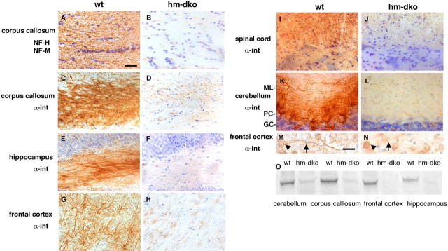
Alpha-internexin, a neuronal intermediate filament protein implicated in neurodegenerative disease, coexists with the neurofilament (NF) triplet proteins (NF-L, NF-M, and NF-H) but has an unknown function. The earlier peak expression of alpha-internexin than the triplet during brain development and its ability to form homopolymers, unlike the triplet, which are obligate heteropolymers, have supported a widely held view that alpha-internexin and neurofilament triplet form separate filament systems. Here, we demonstrate, however, that despite a postnatal decline in expression, alpha-internexin is as abundant as the triplet in the adult CNS and exists in a relatively fixed stoichiometry with these subunits. Alpha-internexin exhibits transport and turnover rates identical to those of triplet proteins in optic axons and colocalizes with NF-M on single neurofilaments by immunogold electron microscopy. Alpha-internexin also coassembles with all three neurofilament proteins into a single network of filaments in quadruple-transfected SW13vim(-) cells. Genetically deleting NF-M alone or together with NF-H in mice dramatically reduces alpha-internexin transport and content in axons throughout the CNS. Moreover, deleting alpha-internexin potentiates the effects of NF-M deletion on NF-H and NF-L transport. Finally, overexpressing a NF-H-LacZ fusion protein in mice induces alpha-internexin and neurofilament triplet to aggregate in neuronal perikarya and greatly reduces their transport and content selectively in axons. Our data show that alpha-internexin and the neurofilament proteins are functionally interdependent. The results strongly support the view that alpha-internexin is a fourth subunit of neurofilaments in the adult CNS, providing a basis for its close relationship with neurofilaments in CNS diseases associated with neurofilament accumulation.


Figures

Figure 1.
Figure 1.
Immunologic identification of α-internexin in Triton-soluble and Triton-insoluble fractions of optic axon separated by two-dimensional gel electrophoresis. A, B, Only the relevant portions of the 2D gels are depicted. C–E, In mouse axons, α-internexin is a 58 kDa protein with an isolectric point of 5.5–5.6 (C) and is exclusively Triton-insoluble and heavily radiolabeled in optic axons after intravitreal injection of 35S-methionine (D, E). F–I, α-Internexin and its radiolabeled counterpart are detected in axons (F, G) but not in glial cells of optic nerves (H, I) by immunoblot analyses after selectively labeling axons and glia (see Materials and Methods). B and C are Coomassie blue-stained gels of axonal cytoskeleton (prepared from mouse spinal cord) and optic axons, respectively. D and E are Coomassie blue-stained gel of Triton-insoluble fraction and its corresponding autoradiograph, respectively. F and H are autoradiographs of optic axons and glial cells, respectively; G and I are their corresponding immunoblots stained for α-internexin, respectively. J–L show α-internexin and NF-L ratio along optic pathway. K, L, Coomassie-stained 2D gels of Triton-insoluble fractions from consecutive 1 mm segments of the optic nerve and tract demonstrate that α-internexin and NF-L show the same proximal to distal increase (approximate threefold) (K) and maintain a constant ratio (∼50%) along optic pathway (L). The even numbers in J represent nerve segment numbers. α-Internexin and NF-L are indicated by an arrowhead and arrow, respectively. The asterisk in I is a degradation product of α-internexin. CB, Coomassie blue; S35, autoradiograph; IB, immunoblot.
Figure 2.
Figure 2.
α-Internexin (arrowhead) and NF-L (arrow) have identical axonal transport rates and maintain a constant ratio during transport. A, B, 2D PAGE analysis of consecutive 1 mm segments of the optic nerve and tract were performed on mice at intervals 3–45 d after injecting 35S-methionine (A, 3 d; B, 15 d). C, Regression analysis of replicate series of gels indicated rates of movement of 0.171 ± 0.048 mm/d (mean ± SD; n = 49) for α-internexin and NF-L and 0.105 ± 0.051 mm/d (mean ± SD; n = 49) for Triton-soluble tubulin. D, The ratio of labeled α-internexin to labeled NF-L was constant during intervals 3–45 d after isotope injection.
Figure 3.
Figure 3.
Ultrastructural colocalization of α-internexin and NF-M on the same neurofilament in optic nerve. Paraformaldehyde-fixed samples were incubated with mouse anti-α-internexin and rabbit anti-NF-M mixture (1:5/1:200) and probed with goat anti-mouse IgG and/or goat anti-rabbit IgG conjugated to 10 nm and 5 nm gold beads. A, B, As expected, for the immunodetection of α-internexin in normal mice (A), large numbers of 10 nm gold particles are aligned with most 10 nm filaments in the axon, and negligible numbers are detected in α-internexin knock-out mice (B). C, Decoration of a single filament by 10 nm gold particles conjugated to anti-α-internexin antibody. D, Linear arrays of two sizes of gold particles (10 nm for α-internexin and 5 nm for NFM) decorate most 10 nm filaments in the axon. E, Higher magnification shows that gold particles of two sizes overlie a single filament in the background. Arrowheads point to 10 nm particles (α-internexin), and arrows point to 5 nm particles (NF-M). Scale bars: A, B, D, 200 nm; C, E, 10 nm.
Figure 4.
Figure 4.
Coassembly of α-internexin with all three neurofilament proteins into single filament network. SW13vim(−) cells were quadruple-transfected with constructs that expressed α-internexin, NF-L, NF-M, and NF-H and were immunostained with pairs of antibodies. Each set of three panels across represents the single immunolabels and the merged double label (yellow indicating colocalization) for the following pairs of antibodies: A1–3 and a1–3, rabbit polyclonal antibodies (pAb) to NF-L and mouse monoclonal antibodies (mAb) to NF-M; B1–3 and b1–3, rabbit pAb to NF-L and mouse mAb to NF-H; C1–3 and c1–3, rabbit pAb to NF-L and mouse mAb to α-internexin; D1–3 and d1–3, rabbit pAb to NF-M and mouse mAb to α-internexin; E1–3 and e1–3, rabbit pAb to NF-H and mouse mAb to α-internexin. Scale bar, 20 μm.
Figure 5.
Figure 5.
Effects of NF-M deletion on transport rates and levels of α-internexin, NF-L, and NF-H in optic axons. A, B, Mouse genomic DNA was screened for targeted disruption of NF-M homozygous mice through genotyping the NF-M loci by hybridizing with a sequence from NF-M. C–F, On immunoblots of 10–20 μg of total optic nerve protein extract from 3- to 4-month-old mice, α-internexin and neurofilament triplet subunits were identified with mAbs to α-internexin, NF-M, NF-H, and NF-L (C, D, E, and F, respectively), relative levels of each protein were determined by NIH imaging. Measurements are mean ± SD from four animals for each group. In the absence of NF-M protein (D), the levels of α-internexin, NF-H, and NF-L proteins were 54 ± 8% (C), 33 ± 8% (E), and 22 ± 3% (F) of the wild-type level, respectively. G–I, Slow axonal transport analyses, after intravitreal injection of 35S-methionine in control (G) and mko mice (H), demonstrate increased velocities of α-internexin, NF-H, and NF-L and unchanged rates of Triton-insoluble tubulin and actin in mko mice (I). J, Distribution of α-internexin, NF-H, and NF-L in mko along optic pathway compared with that in wild-type mice. The squares represent wild-type control, and diamonds represent knock-out mice.
Figure 6.
Figure 6.
Effects of combined deletion of NF-H and NF-M on α-internexin and NF-L transport and levels in optic axons. hm-dko mice were generated by crossbreeding and screening. A–D, Mouse genomic DNA was screened for targeted disruption of both NF-H (A, B) and NF-M (C, D) homozygous mice through genotyping the NF-H and NF-M loci by hybridizing with a sequence from NF-H or NF-M. NF-H, NF-M, and α-internexin (α-int) were identified by immunoblots of total optic nerve protein (10–20 μg) from 3- to 4-month-old mice using mAbs to NF-H (N52), NF-M (NN18), and α-internexin (MAB5224), respectively. E–G, In mice lacking NF-H and NF-M (E, F), α-internexin levels are <10% of wild-type (wt) levels detected by immunoblotting (G), and NF-L proteins are barely detectable as shown previously (Yuan et al., 2003). H, I, Slow axonal transport analyses (7 d after injection) in wild-type control (H) and hm-dko mice (I), as in Figure 5, reveal barely detectable transport of α-internexin in hm-dko axons compared with wild-type mice. The positions of cytoskeletal proteins are indicated, and the nerve segments are numbered below the gels consecutively from the level of the eye. J, K, 2D transport analyses (7 d after injection) confirm dramatically reduced transport of α-internexin into hm-dko axons. The asterisks in J and K indicate Hsc70.
Figure 7.
Figure 7.
α-Internexin depletion from CNS axons in NF-H and NF-M double-deleted mice. Immunoactivity of α-internexin was detected with MAB5224 (C–N) and of NF-H and NF-M (A, B) detected with SMI33. A, B, NF-H and NF-M were absent in hm-dko mice as shown in corpus callosum. C–L, α-Internexin was markedly depleted from axons in all CNS regions, including corpus callosum (C, D), the polymorph layer of hippocampus (E, F), frontal cortex (G, H), spinal cord (I, J), and cerebellum (K, L). M, N, At higher magnification, α-internexin immunolabeling of cortical neurons includes cell bodies (arrowheads) and neurites (arrows) in wild-type mice (M); but in hm-dko α-internexin, immunolabeling was limited to the cell bodies (N). O, Western blot analysis confirms the marked α-internexin reductions. Scale bars: A–L, 30 μm; M, N, 15 μm. ML, Molecular layer; PC, Purkinje cells; GC, granule cells.
Figure 8.
Figure 8.
Presence of low-level NF-H and NF-M in the parallel fibers of cerebellar granule cells in wild-type (wt) mice. A–D, Immunocytochemistry with SMI 33 and SMI 31 antibodies confirms the presence of low-level NF-H and NF-M in the parallel fibers of cerebellar granule cells in wild-type mice (A, C) and their absence in hm-dko mice (B, D). E–H, Anti-NF-H antibody N52 detects NF-H in the parallel fiber (E) and demonstrates its colocalization with α-internexin (F–H). A and B were stained with SMI33, and C and D were stained with SMI31. E and F were stained with N52, and G was stained with α-internexin antibody. H is the merge of F and G. ML, Molecular layer; PC, Purkinje cells; GC, granule cells. The arrows point to parallel fibers. Scale bars: A, B, 160 μm; C–H, 40 μm.
Figure 9.
Figure 9.
Effects of combined deletion of α-internexin and NF-M on levels and transport of NF-H and NF-L in optic axons. α-Im-dko mice were generated by crossbreeding and screening. A–D, Mouse genomic DNA was screened for targeted disruption of both α-internexin (A, B) and NF-M (C, D) homozygous mice through genotyping the α-internexin and NF-M loci by hybridizing with a sequence from α-internexin or NF-M. E–H, Immunoblot analyses of total optic nerve proteins (10–20 μg) from 3- to 4-month-old mice using mAbs to α-internexin, NF-M, NF-H, and NF-L (E, F, G, and H, respectively) demonstrated the absence of α-internexin and NF-M protein in α-im-dko mice (E, F) and decreased levels of NF-H (27 ± 2%) (G) and NF-L 17 ± 5% (H), respectively, compared with wild-type mice. Measurements are mean ± SD from four animals for each group. Slow axonal transport analyses 14 d after injection indicates undetectable radiolabeled NF-H and NF-L transport even after exposing films for 3 months in α-im-dko mice (J). The appearance of the dark band in J, lane 2, is labeled insoluble tubulin because of long-time (90 d) exposure in the mutant (H) versus short-time (10 d) exposure in the wild-type control (I).
Figure 10.
Figure 10.
Effects of combined α-internexin and NF-H deletion on the transport of NF-L and NF-M in optic axons. α-Ih-dko mice were generated by crossbreeding and screening. A, B, Mouse genomic DNA was screened for targeted disruption of both α-internexin (A) and NF-H (B) homozygous mice through genotyping the α-internexin and NF-H loci by hybridizing with a sequence from α-internexin or NF-H. C–F, Immunoblot analyses of optic nerve proteins (10–20 μg) from 3- to 4-month-old mice with mAbs to α-internexin, NF-H, NF-M, and NF-L (C, D, E, and F, respectively) demonstrates the absence of α-internexin (C) and NF-H protein in α-ih-dko mice (D) but unchanged levels of NF-M (E) and NF-L (F). Measurements are mean ± SD from four animals for each group. G–I, Slow axonal transport analyses (14 d after injection) revealed increased velocity (I) of NF-L and NF-M transport in α-ih-dko mice (H) compared with control mice (G). Note the increased velocity of NF-L and NF-M transport mainly at the location of neurofilament bulk but not the leading edge in α-ih-dko mice. The squares represent wild-type control, and diamonds are knock-out mice.
Figure 11.
Figure 11.
α-Internexin and NF-L transport and levels in optic axons and in retinas containing retinal ganglion cell bodies in NF-H-LacZ transgenic mice. A–D, Quantification image analysis of Coomassie blue-stained 2D gels indicates that α-internexin (arrowheads) and NF-L (arrows) in NF-H-LacZ transgenic mice were <25% of wild-type levels, whereas insoluble tubulin, actin, and spectrin were not changed. E, F, Levels of α-internexin and NF-L in the retina containing retinal ganglion cell bodies are increased in NF-H-LacZ transgenic mice compared with control mice.

Similar articles

Cited by

References

    1. Al-Chalabi A, Miller CC. Neurofilaments and neurological disease. BioEssays. 2003;25:346–355. - PubMed
    1. Archer DR, Watson DF, Griffin JW. Phosphorylation-dependent immunoreactivity of neurofilaments and the rate of slow axonal transport in the central and peripheral axons of the rat dorsal root ganglion. J Neurochem. 1994;62:1119–1125. - PubMed
    1. Beaulieu JM, Robertson J, Julien JP. Interactions between peripherin and neurofilaments in cultured cells: disruption of peripherin assembly by the NF-M and NF-H subunits. Biochem Cell Biol. 1999;77:41–45. - PubMed
    1. Benson DL, Mandell JW, Shaw G, Banker G. Compartmentation of alpha-internexin and neurofilament triplet proteins in cultured hippocampal neurons. J Neurocytol. 1996;25:181–196. - PubMed
    1. Brown A. Visualization of single neurofilaments by immunofluorescence microscopy of splayed axonal cytoskeletons. Cell Motil Cytoskeleton. 1997;38:133–145. - PubMed

Publication types

MeSH terms

LinkOut - more resources