. 2006 Sep 27;26(39):10068-78.
doi: 10.1523/JNEUROSCI.0896-06.2006.

Cytosolic catechols inhibit alpha-synuclein aggregation and facilitate the formation of intracellular soluble oligomeric intermediates

Affiliations

Cytosolic catechols inhibit alpha-synuclein aggregation and facilitate the formation of intracellular soluble oligomeric intermediates

Joseph R Mazzulli et al. J Neurosci. .

Abstract

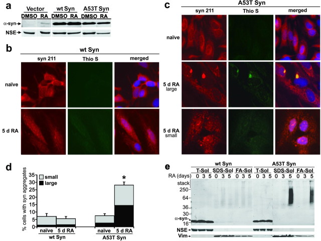
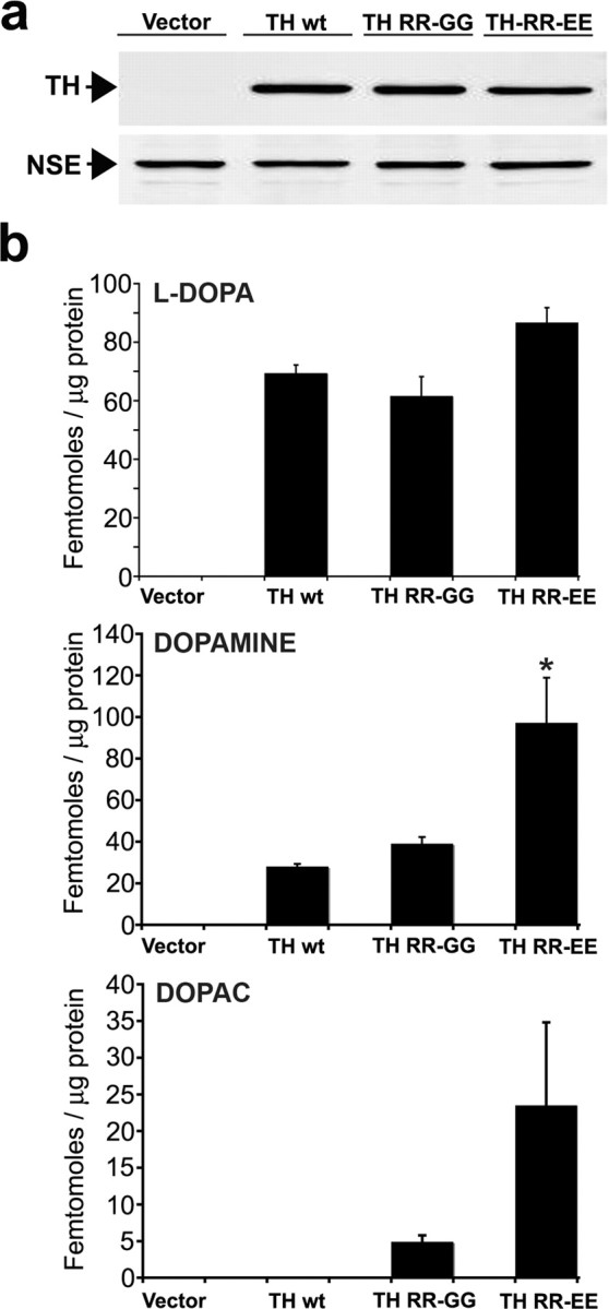
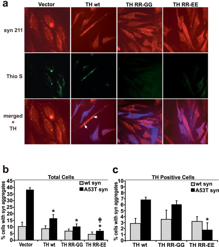
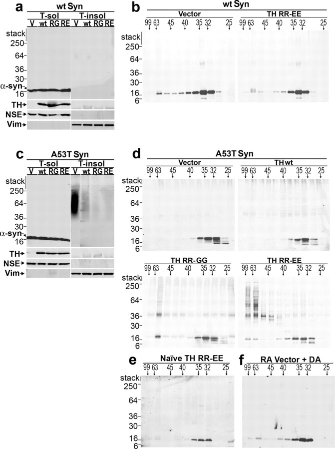
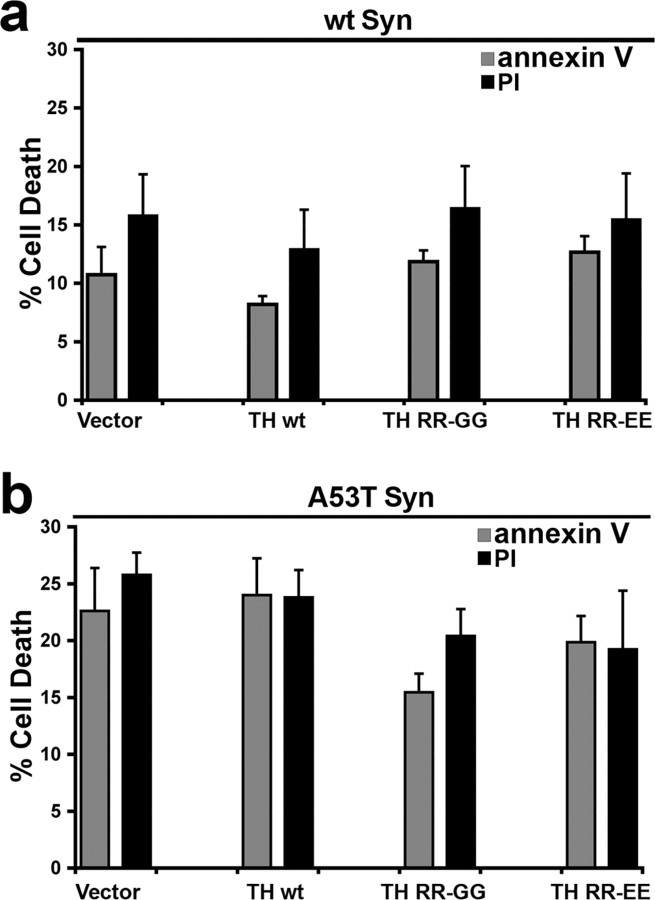
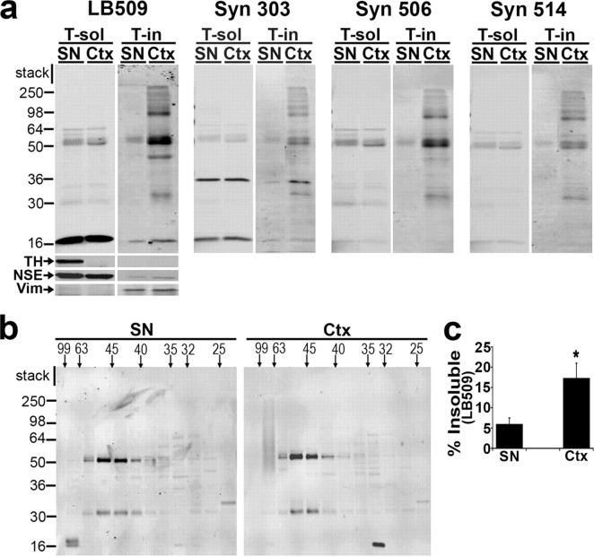
Aberrant aggregation of alpha-synuclein (alpha-syn) to form fibrils and insoluble aggregates has been implicated in the pathogenic processes of many neurodegenerative diseases. Despite the dramatic effects of dopamine in inhibiting the formation of alpha-syn fibrils by stabilization of oligomeric intermediates in cell-free systems, no studies have examined the effects of intracellular dopamine on alpha-syn aggregation. To study this process and its association with neurodegeneration, intracellular catechol levels were increased to various levels by expressing different forms of tyrosine hydroxylase, in cells induced to form alpha-syn aggregates. The increase in the steady-state dopamine levels inhibited the formation of alpha-syn aggregates and induced the formation of innocuous oligomeric intermediates. Analysis of transgenic mice expressing the disease-associated A53T mutant alpha-syn revealed the presence of oligomeric alpha-syn in nondegenerating dopaminergic neurons that do contain insoluble alpha-syn. These data indicate that intraneuronal dopamine levels can be a major modulator of alpha-syn aggregation and inclusion formation, with important implications on the selective degeneration of these neurons in Parkinson's disease.


Figures

Figure 1.
Figure 1.
Differentiation of SH-SY5Y cells expressing A53T α-syn induces the formation of amyloidogenic, Triton-insoluble aggregates. SH-SY5Y cells were stably transfected with empty vector, wt Syn, or A53T α-syn (A53T Syn) expression plasmids and cultured with or without 20 μm RA. a, Cells were harvested after 5 d of culture with either DMSO (vehicle) or 20 μm RA. Triton-soluble lysates (40 μg of protein/lane) were analyzed by Western blot analysis using monoclonal antibody Syn 211, and NSE was used as a loading control. b, wt α-syn cells were stained with the anti-α-syn monoclonal antibody Syn 211 (red) and 0.05% thioflavin S to detect amyloidogenic aggregates (Thio S; green) (20× magnification). The majority of naive cells show a diffuse staining pattern for α-syn, and differentiation by RA treatment for 5 d does not change the distribution patterns of α-syn. c, A53T α-syn cells were analyzed as in b, showing the induction of α-syn aggregation by RA differentiation. Both large juxtanuclear aggregates (middle row) and small punctate aggregates (bottom row) were observed. d, Quantification of the number of cells containing α-syn-positive punctate structures. Values are the mean ± SEM (n = 3). *p = 0.01 for total aggregates relative to A53T α-syn naive condition. e, wt or A53T α-syn cells were subjected to sequential biochemical extraction in 1% Triton-X (T-sol), 2% SDS (SDS-sol), and 70% formic acid (FA-sol), followed by Western blot analysis using LB509. Similar results were obtained when the blot was stripped and reprobed with Syn 211. To monitor the efficiency of the extractions and protein loading, NSE was used for Triton-soluble fractions, whereas the Triton-insoluble intermediate filament protein vimentin (Vim) was used for SDS- and FA-soluble fractions. The molecular weight marker located at the left side of the blot indicates the protein size in kilodaltons. Although differentiated A53T α-syn cells contain Triton-insoluble α-syn, wt-syn cells do not form detectable amounts of Triton-insoluble materials after 5 d in culture.
Figure 2.
Figure 2.
Manipulation of intracellular catechol levels in SH-SY5Y cells expressing A53T α-syn by infection with lentivirus-containing TH expression plasmids. A53T α-syn-expressing cells were infected with lentivirus generated with empty vector plasmid (Vector), or vector expressing wt TH (TH wt), 37,38 RR-GG TH (TH RR-GG), or 37,38 RR-EE TH (TH RR-EE). The cells were infected using a multiplicity of infection that transduced ∼50% of total cells and cultured for 5 d in the presence of 20 μm RA. a, Western blot analysis of TH-infected cells shows similar expression of each TH protein. NSE was used as a loading control. b, Quantification of intracellular catechol levels HPLC with on-line electrochemical detection. Similar results were obtained when wt α-syn-expressing cells were infected with TH-containing plasmids (data not shown). Values are the mean ± SEM (n = 3). *p < 0.05 relative to TH wt and TH RR-GG.
Figure 3.
Figure 3.
Increasing TH protein expression and intracellular catechol levels decreases the number of α-syn aggregates in A53T α-syn-expressing cells. wt α-syn- and A53T α-syn-expressing cells were infected with lentivirus containing TH expression plasmids and cultured for 5 d in 20 μm RA. a, A53T α-syn cells were stained with Syn 211 (red), 0.05% thioflavin S (Thio S; green) to visualize amyloidogenic aggregates, and anti-TH antibodies (blue) (20× magnification). The arrows show that thioflavin S-positive structures were also detected in the cells treated with TH lentivirus, although this was observed mostly in cells that had not been transduced. b, Quantification of total cells containing α-syn punctate structures using both LB509 (n = 2) and Syn 211 (n = 3) antibodies. Values are the mean ± SEM (n = 5). *p < 0.05 relative to vector control; p < 0.05 relative to TH wt. c, Quantification of only the TH-positive cells containing α-syn punctate structures using LB509 and Syn 211 shows that the observed decrease in α-syn aggregates correlates with increased catechol production by the TH RR-EE mutant rather than TH protein levels. Values are the mean ± SEM (n = 3). *p < 0.05 relative to TH wt and p < 0.05 relative to TH RR-GG.
Figure 4.
Figure 4.
Increasing intracellular catechol levels decreases Triton-insoluble A53T α-syn with a concomitant accumulation of soluble oligomers. a, wt α-syn-expressing SH-SY5Y cells were transduced with lentivirus constructed with the empty vector or lentivirus containing wt, RR-GG, or RR-EE TH expression plasmids and cultured for 5 d in the presence of 20 μm RA. Cell lysates were subjected to sequential extraction and analyzed by Western blot using LB509. Western blot for TH verifies similar expression of each TH variant. NSE was used to monitor extraction and loading for Triton-soluble fractions, and vimentin was used for Triton-insoluble fractions. b, Triton-soluble fractions from wt α-syn cells were analyzed by SEC using a superdex 200 HR 10/30 gel filtration column. Fractions were collected and analyzed by Western blot analysis using antibody Syn 211. The horizontal marker indicates the molecular size in angstroms, whereas the vertical marker indicates the molecular weight in kilodaltons. A value of 99 Å corresponds to the column void volume (∼2000 kDa), and the α-syn monomer elutes off at a peak between 32 and 35 Å (or ∼57 kDa). c, A53T α-syn cells were infected, differentiated, and analyzed as in a. d, Triton-soluble fractions from A53T α-syn cells were analyzed by SEC as in b. SDS/heat-stable dimers, trimers, and oligomers are not detected in 50 μg of total cell protein (c) but were detected when 1 mg of total cell protein is analyzed by SEC. e, Naive A53T α-syn-expressing cells were infected with lentivirus containing TH RR-EE plasmid and cultured without RA for 5 d. SEC/Western blot analysis of the Triton-soluble fraction using antibody Syn 211 reveals the absence of α-syn eluting at the void volume. f, A53T α-syn cells were infected with lentivirus containing empty vector plasmid and cultured for 5 d with RA. The addition of 0.5 μm DA in the lysis buffer (threefold excess of the concentration detected in TH RR-EE-infected cells) did not dramatically shift the elution profile of α-syn, suggesting that catechol-induced oligomers are formed by active intracellular processes. The data are representative of three independent experiments. V, Vector; RG, RR-GG; RE, RR-EE; Vim, vimentin; T-sol, Triton-soluble fractions; T-insol, Triton-insoluble fractions.
Figure 5.
Figure 5.
Accumulation of catechol-induced α-syn oligomers does not accelerate A53T α-syn-mediated cell death in SH-SY5Y cells. wt and A53T α-syn cells were infected with lentivirus generated with empty vector or TH-containing plasmids, and the percentage of FITC-conjugated annexin V- and PI-positive cells was measured after 5 d in culture with RA by flow cytometry. Although A53T α-syn-expressing cells degenerate more rapidly compared with wt α-syn cells, increasing catechol levels does not change cell viability in either wt or A53T α-syn-expressing cell lines. Values are the mean ± SEM (n = 4–6). Similar results were obtained when measuring cell viability by trypan blue exclusion (data not shown).
Figure 6.
Figure 6.
Decreased Triton-insoluble α-syn and an increased proportion of 63–99 Å α-syn oligomers in nondegenerating SN of diseased A53T tg mice. a, Ctx and SN from A53T mice displaying motor impairments were dissected, sequentially extracted (see Materials and Methods), and analyzed by Western blot using a panel of α-syn antibodies (50 μg of total protein/lane). SN tissue contains TH as expected, whereas it is not detected in the Ctx. Western blot for NSE was used to monitor extraction and loading for Triton-soluble (T-sol) fractions, whereas vimentin (Vim) was used for Triton-insoluble (T-in) fractions. The molecular weight marker indicates the protein size in kilodaltons. b, T-soluble fractions were analyzed by SEC, followed by Western blot using the monoclonal antibody Syn 211 (500 μg of total protein injected on column). The horizontal marker indicates the molecular size in angstroms. c, The level of Triton-insoluble α-syn detected by LB509 was quantified by densitometry and normalized to vimentin (percentage of insoluble α-syn: SN, 6 ± 1; Ctx, 17 ± 3). Values are the mean ± SEM (n = 3). *p < 0.05. Although Ctx tissue contains α-syn-positive perikaryal inclusions and dystrophic neurites, cells of the SN are completely devoid of α-syn pathology in this mouse model (Giasson et al., 2002).

Similar articles

Cited by

References

    1. Abeliovich A, Schmitz Y, Farinas I, Choi-Lundberg D, Ho WH, Catillo PE, Shinsky N, Verdugo JM, Armanini M, Ryan A, Hynes M, Phillips H, Sulzer D, Rosenthal A. Mice lacking alpha-synuclein display functional deficits in the nigrostriatal dopamine system. Neuron. 2000;25:239–252. - PubMed
    1. Ara J, Przedborski S, Naini AB, Jackson-Lewis V, Trifiletti RR, Horwitz J, Ischiropoulos H. Inactivation of tyrosine hydroxylase by nitration following exposure to peroxynitrite and 1-methyl-4-phenyl-1,2,3,6-tetrahydropyridine (MPTP) Proc Natl Acad Sci USA. 1998;95:7659–7663. - PMC - PubMed
    1. Baba M, Nakajo S, Tu P, Tomita T, Nakaya K, Lee VM, Trojanowski JQ, Iwatsubo T. Aggregation of alpha-synuclein in Lewy bodies of sporadic Parkinson's disease and dementia with Lewy bodies. Am J Pathol. 1998;152:879–884. - PMC - PubMed
    1. Cabin DE, Shimazu K, Murphy DD, Cole NB, Gottschalk W, McIIwain KL, Orrison B, Chen A, Ellis CE, Paylor R, Lu B, Nussbaum RL. Synaptic vesicle depletion correlates with attenuated synaptic responses to prolonged repetitive stimulation in mice lacking α-synuclein. J Neurosci. 2002;22:8797–8807. - PMC - PubMed
    1. Cappai R, Leck SL, Tew DJ, Williamson NA, Smith DP, Galatis D, Sharples RA, Curtain CC, Ali FE, Cherny RA, Culvenor JG, Bottomley SP, Masters CL, Barnham KJ, Hill AF. Dopamine promotes alpha-synuclein aggregation into SDS-resistant soluble oligomers via a distinct folding pathway. FASEB J. 2005;19:1377–1379. - PubMed

Publication types

MeSH terms

LinkOut - more resources