. 2006 Dec;17(12):5241-52.
doi: 10.1091/mbc.e06-07-0634. Epub 2006 Sep 27.

Rendezvin: An essential gene encoding independent, differentially secreted egg proteins that organize the fertilization envelope proteome after self-association

Affiliations

Rendezvin: An essential gene encoding independent, differentially secreted egg proteins that organize the fertilization envelope proteome after self-association

Julian L Wong et al. Mol Biol Cell. 2006 Dec.

Abstract

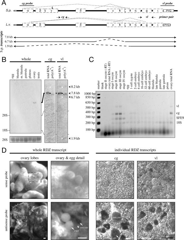
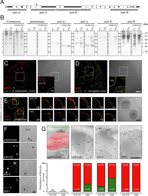
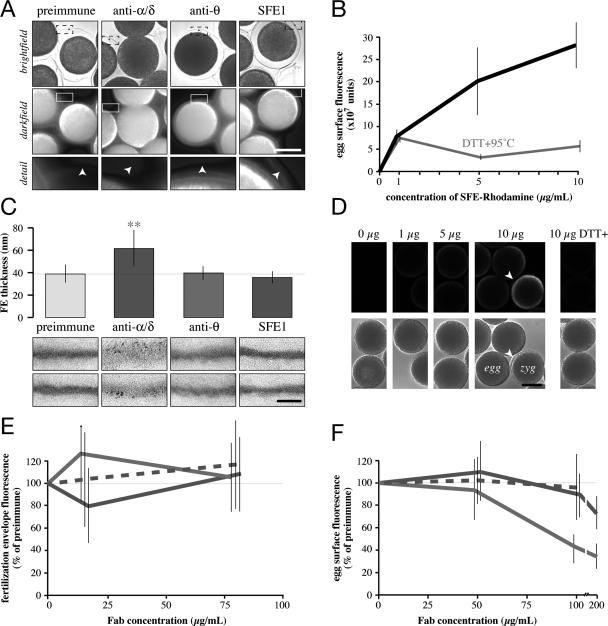
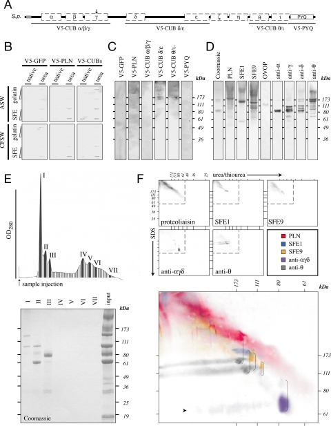
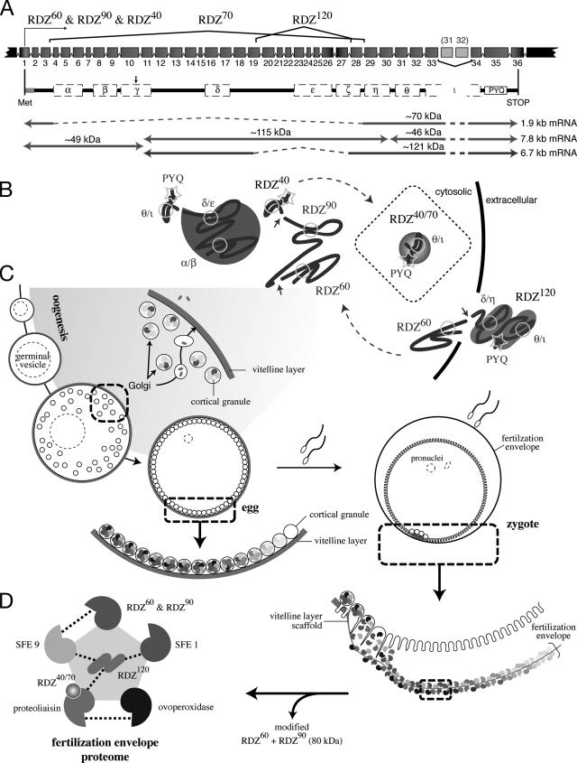
Preventing polyspermy during animal fertilization relies on modifications to the egg's extracellular matrix. On fertilization in sea urchins, the contents of cortical granules are secreted and rapidly assemble into the egg's extracellular vitelline layer, forming the fertilization envelope, a proteinaceous structure that protects the zygote from subsequent sperm. Here, we document rendezvin, a gene whose transcript is differentially spliced to yield proteins destined for either cortical granules or the vitelline layer. These distinctly trafficked variants reunite after cortical granule secretion at fertilization. Together, they help coordinate assembly of the functional fertilization envelope, whose proteome is now defined in full.


Figures

Figure 1.
Figure 1.
Rendezvin transcripts are exclusive to oocytes. (A, top) Schematic identifying splicing organization predicted by RT-PCR fragments in Figure 2 (dashed lines), probe locations (S. purpuratus), and primer pairs (L. variegatus) relative to the predicted open reading frames, including motifs (see Supplemental Figure 1). cg, cortical granule; vl, vitelline layer. (A, bottom) Diagram of S. purpuratus coding strand RNA identified by sequenced RT-PCR products, listed with likely size of each transcript based on RNA blot pattern. (B) RNA blots probing S. purpuratus total RNA (10 μg) from different developmental stages and gonads, or S. purpuratus ovary total RNA (2.5 μg) and oocyte-enriched poly-A+ RNA (1 μg). The developmental blot (left) was probed with an equal mixture of antisense RNA probes. Duplicate RNA blots (right) were probed with either rdz probe and then overexposed to identify all major transcripts (white dots). Approximate size of each identified transcript is also shown, calculated based on migration distance (our unpublished data). rRNA bands are indicated. Ethidium bromide staining for the 28S rRNA bands is shown as loading controls for the developmental blot. (C) Ethidium bromide image of multiplex reverse transcriptase PCR amplifications from two-cell/embryo equivalents of whole L. variegatus cells or embryos, or 250 ng of ovary total RNA. Primer pairs for the vitelline layer or cortical granule transcript of rendezvin, the cortical granule protein SFE9, and the 18S rRNA (Supplemental Table 3) were used to amplify specific products from total lysates of each stage indicated. Oocyte staging was based on the relative nuclear-to-cytoplasmic ratio, as described previously (Wong et al., 2004). Controls include no template (“blank”) and reactions without reverse transcriptase (“-RT”). (D) In situ hybridizations of rendezvin on S. purpuratus ovary and eggs. Left set shows intact ovary lobes and ovulated eggs, including a magnified image. Right set shows tissue smears of ovary hybridized with either probe (see A). Both sense (top images) and antisense (bottom images) are shown. Oocytes (oo), their germinal vesicles (gv), and eggs are labeled. Bars, 100 μm.
Figure 2.
Figure 2.
Rendezvin variants localize to distinct organelles. (A) Schematic diagram of the S. purpuratus rendezvin open reading frame indicating regions used to generate specifically named antisera. Diagram includes the structural motifs of orthologues (Supplemental Figure 1). Note polyclonal anti-RDZ or anti-SFE1 sera, for a cortical granule protein (Wessel et al., 2000), were used as a control in labeling and transmission electron micrograph localization. (B) Comparison of contents of S. purpuratus surface complex (“sc”; 25 μg per lane) and FE (5 μg), stained for total protein (Coomassie), or probed with each serum. Samples were separated under nonreducing (left) and reducing (right) SDS-PAGE. Pattern from each antiserum (see A) compared with a mix of their respective preimmune serum. (C and D) Single confocal images of ovary sections from S. purpuratus (C) and L. variegatus (D) probed with anti-SFE1 (red) and anti-θ (green). Fluorescence signal is indicated on the left panel with respective differential interference contrast (DIC) images found in the right panels. Details (insets) are shown, with boxes indicating their origin: oocyte (oo) and eggs (egg) are outlined. Bar, 50 μm. (E) Permeabilized, stratified L. variegatus oocyte (oocyte) and egg (egg) double-stained for SFE1 (red) and anti-θ (green). Two-dimensional projection of optical sections (images 13–19) through an equatorial region of each cell is shown (left) with corresponding differential interference contrast image (right). Details of centrifugal poles per cell are shown as independent optical sections (middle panels). Z-distance between each optical section is 2.5 μm. Bar, 50 μm. (F) Single antibody staining of egg cortical lawns. Confocal fluorescence (2 airy units, left) and DIC (right) images are shown. Free cortical granules (cg) are indicated (arrowhead). Note membrane on lawns that have folded over and are exposed. Bar, 5 μm. (G) Transmission electron micrographs of immunogold localizations on S. purpuratus oocytes probed with anti-αγδ, anti-θ, or anti-SFE1. Quantitation of colloidal gold particle distribution, as percentage of total particles counted over 15–20 sections of oocytes or eggs. Percentiles refer to the cortical granule group; number of particles scored per treatment is indicated (n). Preimmune serum for anti-θ is shown, with colorization to identify the organelles analyzed in particle scoring. Arrowhead emphasizes particles in the vitelline layer. Bar, 500 nm.
Figure 3.
Figure 3.
Rendezvin components play distinct and essential roles in the fertilization envelope. (A) Analysis of FE birefringence, comparing bright-field (top) and darkfield (middle; composite of thresholded pseudocolor images emphasizing the intensity of light derived from the FE overlaying grey scale darkfield image) images of eggs activated by ionophore in the presence of antisera (1:20 dilution). Darkfield details of FEs (bottom) from one segment of the larger image (box) are shown, with arrowheads indicating the FE as seen by bright-field. Bar, 50 μm. (C) Quantitation of the thickness of respective FEs formed in the presence of 1:20 dilutions of antisera, with representative electron micrographs of each FE (bottom). Vertical bars indicate SD. **p < 10−9 among all treatments. Bar, 500 nm. (E) Competition of soluble, biotinylated S. purpuratus FE proteins to intact FEs in the presence of increasing concentrations of mixed Fabs. The x-axis is shown logarithmically. Vertical lines indicate SD. Color coordinated to C. (B) Titration of native (black) or denatured (gray) S. purpuratus rhodamine-FE binding to eggs (see Materials and Methods). Vertical lines indicate SD. (D) Representative confocal images showing dose-dependent binding of rhodamine-FE to the egg surface. Arrowhead identifies a partial FE showing higher binding intensity, consistent with results from E. Bar, 50 μm. (F) Competition of rhodamine-FE binding to the egg by purified Fabs. Color coordinated to C. Vertical lines indicate SD.
Figure 4.
Figure 4.
Rendezvin is integral to fertilization envelope assembly. (A) Schematic diagram identifying regions of S. purpuratus rendezvin used to generate V5-recombinant protein. The amino-terminal sequence of S. purpuratus PLN, previously reported to interact with other FE proteins (Somers and Shapiro, 1991), was also used. (B and C) V5-recombinants were tested for binding to immobilized FEs (see Materials and Methods). (B) One and 5 μg of urea-denatured gelatin or FE was probed with a total of 5 μg of V5-recombinant protein in seawater containing (ASW) or lacking (CFSW) free calcium. (C) Proteins separated by urea-PAGE were probed with a total of 10 μg of V5-reactive equivalents of individual or paired V5-recombinants in CFSW. (D) One microgram of FE was separated into subcomplexes by urea-PAGE and immunoblotted for individual proteins or domains of RDZ to identify coassociating proteins. (E) UV absorption profile and eluting fractions of 10 μg of total FE separated by gel filtration identify seven major protein peaks (top). Only the first three peaks contain significant protein as detected by SDS-PAGE and Coomassie staining (bottom). (F) Two micrograms of FE were separated by two-dimensional urea-SDS-PAGE and immunoblotted for the major structural proteins found within the fertilization envelope (top, gray). Details superimposing all the immunoblots (bottom, color) indicate structural proteins that interact endogenously in a pairwise manner (brackets). The faint streak of a 70-kDa protein is indicated (arrowhead).
Figure 5.
Figure 5.
Model of rendezvin protein expression and the fertilization envelope proteome. (A) Schematic of the S. purpuratus rendezvin exons, colored to match the predicted protein encoded, and resultant transcripts following posttranscriptional processing. The duplication of exons 31 (185 base pairs) and 32 (191 base pairs) is not retained in the most stable rdz transcript (8.2 kb versus stable 7.8 kb; our unpublished data). Spliced regions are shown with the corresponding isoform nomenclature used. Predicted primary structure, after removal of exons (dashed lines) and proteolysis (arrow; see text), are shown below with their predicted protein mass. The approximate transcript lengths that correspond to each isoform are listed on the right. Note the amino-terminal isoform (RDZ60; ∼49 kDa) is shared by two transcripts due to proteolysis. (B) Sketches of possible secondary structures for each RDZ component predicted by splice variants and immunoblot detection, including the interacting CUBs, are shown. (C) Diagram of oogenesis and fertilization, including a model for RDZ translation and protein trafficking within the oocyte (shaded region). Includes details of the egg and zygotic cortex undergoing exocytosis. (D) One binding network of interacting structural protein partners within the fertilization envelope is shown, following the color representation of Figure 4F.

Similar articles

Cited by

References

    1. Andersen O. M., Christensen L. L., Christensen P. A., Sorensen E. S., Jacobsen C., Moestrup S. K., Etzerodt M., Thogersen H. C. Identification of the minimal functional unit in the low density lipoprotein receptor-related protein for binding the receptor-associated protein (RAP). A conserved acidic residue in the complement-type repeats is important for recognition of RAP. J. Biol. Chem. 2000;275:21017–21024. - PubMed
    1. Anderson E. Oocyte differentiation in the sea urchin, Arbacia punctulata, with particular reference to the origin of cortical granules and their participation in the cortical reaction. J. Cell Biol. 1968;37:514–539. - PMC - PubMed
    1. Bisgrove B. W., Raff R. A. The SpEGF III gene encodes a member of the fibropellins: EGF repeat-containing proteins that form the apical lamina of the sea urchin embryo. Dev. Biol. 1993;157:526–538. - PubMed
    1. Bork P., Beckmann G. The CUB domain. A widespread module in developmentally regulated proteins. J. Mol. Biol. 1993;231:539–545. - PubMed
    1. Bruskin A. M., Tyner A. L., Wells D. E., Showman R. M., Klein W. H. Accumulation in embryogenesis of five mRNAs enriched in the ectoderm of the sea urchin pluteus. Dev. Biol. 1981;87:308–318. - PubMed

LinkOut - more resources