. 2006 Dec;17(12):5185-97.
doi: 10.1091/mbc.e06-05-0469. Epub 2006 Sep 27.

TIF1 activates the intra-S-phase checkpoint response in the diploid micronucleus and amitotic polyploid macronucleus of Tetrahymena

Affiliations

TIF1 activates the intra-S-phase checkpoint response in the diploid micronucleus and amitotic polyploid macronucleus of Tetrahymena

J Sebastian Yakisich et al. Mol Biol Cell. 2006 Dec.

Abstract

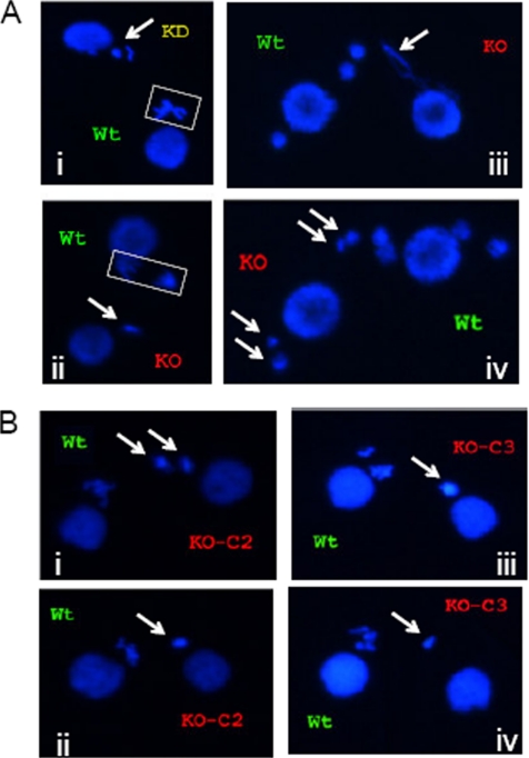
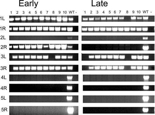
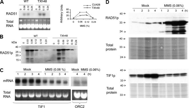
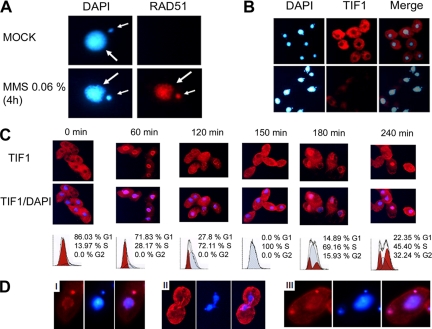
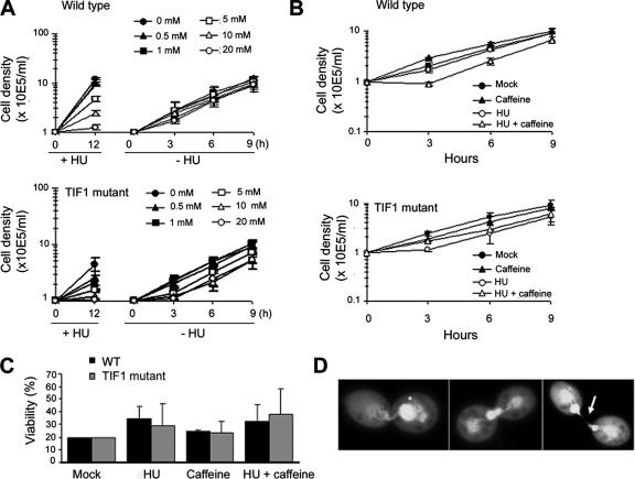
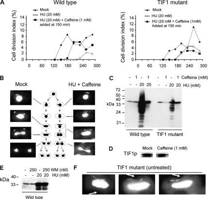
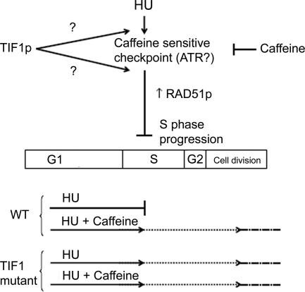
The ribosomal DNA origin binding protein Tif1p regulates the timing of rDNA replication and is required globally for proper S-phase progression and division of the Tetrahymena thermophila macronucleus. Here, we show that Tif1p safeguards chromosomes from DNA damage in the mitotic micronucleus and amitotic macronucleus. TIF1p localization is dynamically regulated as it moves into the micro- and macronucleus during the respective S phases. TIF1 disruption mutants are hypersensitive to hydroxyurea and methylmethanesulfonate, inducers of DNA damage and intra-S-phase checkpoint arrest in all examined eukaryotes. TIF1 mutants incur double-strand breaks in the absence of exogenous genotoxic stress, destabilizing all five micronuclear chromosomes. Wild-type Tetrahymena elicits an intra-S-phase checkpoint response that is induced by hydroxyurea and suppressed by caffeine, an inhibitor of the apical checkpoint kinase ATR/MEC1. In contrast, hydroxyurea-challenged TIF1 mutants fail to arrest in S phase or exhibit caffeine-sensitive Rad51 overexpression, indicating the involvement of TIF1 in checkpoint activation. Although aberrant micro- and macronuclear division occurs in TIF1 mutants and caffeine-treated wild-type cells, TIF1p bears no similarity to ATR or its substrates. We propose that TIF1 and ATR function in the same epistatic pathway to regulate checkpoint responses in the diploid mitotic micronucleus and polyploid amitotic macronucleus.


Figures

Figure 1.
Figure 1.
Cytogenetic evidence for micronuclear genome instability, developmental delay, and/or meiotic arrest in TIF1-deficient strains. The wild-type strain (CU428) was mated with TIF1 mutants (TXh48 [KD, knockdown]; TXk202 [KO, knockout/null]), and mating pairs were examined at various times during development by DAPI fluorescent staining. Wild-type and mutant strains were prelabeled with MitoTracker Red and Green dyes, respectively, to identify each partner. (A) Meiotic aberrations in TIF1-deficient strains. Meiotic stages: i, wild type: metaphase, mutant: prophase (?); ii, wild type: anaphase, mutant: prophase (?); iii, wild type: postmeiotic pronuclei, mutant: meiotic crescent or aberrant anaphase (?); and iv, wild type and mutant: postmeiotic pronuclei. Arrows point to mutant nuclei. (B) Comparative cytogenetic analysis of siblings cells in subcloned parental lines KO-C2 and KO-C3 derived from the tif1-1::neo knockout strain TXh202. The newly generated clonal lines were briefly expanded and then mated with the wild-type strain CU428 to examine meiosis. Arrows point to condensed, micronuclear-derived DAPI-staining chromosomes in siblings within the same mating culture. Micrographs i and ii, representative tif1-1::neo/TIF1 knockout clone C2 siblings; micrographs iii and iv, representative tif1-1::neo knockout clone C3 siblings.
Figure 2.
Figure 2.
Micronuclear genome instability in TIF1-deficient T. thermophila. Ten clonal lines derived from the tif1-1::neo/TIF1 knockdown strain TXh48 were established and subjected to PCR analysis with primers sets that span sites for chromosome breakage sequence (CBS)-mediated chromosome fragmentation in the developing macronucleus. PCR primers derived from the right (R) and left (L) arms of all five micronuclear chromosomes were tested. 1–10, clonal TXh48 knockdown lines; WT, CU428. Early, ∼150 fissions after conferring resistance to high concentrations of pm (encoded by the tif1-1::neo disruption); late, ∼250 fissions later than “early.”
Figure 3.
Figure 3.
Regulation of RAD51 and TIF1 by MMS. (A) RAD51 Northern blot analysis after a 1-h exposure to MMS [0–0.24% (vol/vol)]. Top left, hybridization with a RAD51 coding region probe. Bottom left, ethidium bromide staining of total DNA before transfer to GeneScreen Plus membranes (New England Biolabs, Beverly, MA). Right, graphic representation of RNA hybridization signals quantified on a PhosphorImager (Bio-Rad, Hercules, CA). Dotted line, wild type (CU428); solid line, TIF1 knockdown mutant (TXh48). (B) Western blot analysis of Rad51p in wild-type and mutant whole cell lysates (1-h MMS treatment). (C) Northern blot analysis of TIF1 mRNA in MMS-treated wild-type cells. Strain CU427, same MMS treatment regimen as in C. Probes, TIF1 and origin recognition complex, subunit 2 (ORC2) protein-coding regions. Bottom, ethidium bromide staining of RNA preparations for normalization. (D) Rad51p and Tif1p levels in mock and MMS-treated cells (0.06% MMS; 1–4 h). The Tif1p Western blot probe was directed against the myc epitope in the tif1-2–tagged strain TAM101. Membranes were stained with Ponceau S to compare the amount of total protein loaded in each lane.
Figure 4.
Figure 4.
Immunolocalization of Rad51p and Tif1p in control and MMS-treated cells. (A) Rad51p immunolocalization in wild-type cells (0.06% MMS; 4 h). Red, Rad51p immunofluorescence; blue, DAPI. Small arrow, micronucleus, large arrow, macronucleus. (B) Immunolocalization of Tif1p in asynchronous control and MMS-treated cultures (strain TAM101; 0.06% MMS, 4 h). Red, Tif1p immunofluorescence; blue, DAPI. (C) Cell cycle localization of Tif1p. Strain TAM101 was synchronized by starvation and refeeding and assayed at 30-min intervals for TIF1p localization (red, TIF1p immunofluorescence; blue, DAPI) and DNA content (flow cytometry). Alternating time points are shown. (D) Immunolocalization of Tif1p late in the cell cycle (240 min). Note the micronuclear localization of Tif1p in micrographs i and iii and exclusion from dividing macronuclei in micrograph ii.
Figure 5.
Figure 5.
Response of wild-type and tif1-1::neo mutants to HU and caffeine. (A) Wild-type (CU428) and tif1-1::neo/TIF1 mutant (TXh48) strains were grown to a density of 3 × 10⁴/ml and incubated for 12 h in growth media containing a range of HU concentrations (0–20 mM, +HU; stock solution in water) to induce S-phase–specific cell cycle arrest. Outgrowth of cultures after removal of the drug (−HU), refed cells were adjusted to a density of 1 × 10⁵/ml and counted at 3-h intervals. The mean and SEs for three experiments are shown. (B) Outgrowth of cultures treated for 12 h with 20 mM HU and/or 0.3 mM caffeine (stock solution in water) and released into drug-free media. Cell densities were adjusted to 1 × 10⁵/ml after washing out the drug(s), and the cultures were counted at 3-h intervals. The mean and SE for two experiments are shown. (C) Cell viability analysis in cultures treated with HU and/or caffeine. Wild-type and TIF1-deficient strains were incubated for 12 h in media containing no drug, 0.3 mM caffeine, 20 mM HU, or both. Cells were washed repeatedly to remove the drugs and plated out at decreasing concentrations into two 96-well dishes/dilution. For comparative analysis, the percentage of wells that was positive for growth in mock-treated cultures (plating density 0.3 cells/well) was normalized to 20 for each of the three experiments. The representative data for drug-treated strains were similarly normalized and correspond to the mean and SE of the three experiments. (D) Examples of macronuclear division defects in wild-type cells treated for 12 h with 20 mM HU and 0.3 mM caffeine and then propagated for 7 h in drug-free media.
Figure 6.
Figure 6.
Identification of an intra-S-phase checkpoint defect in Tif1p-deficient T. thermophila. (A) Wild-type (CU428) and tif1-1::neo/ TIF1 mutant (TXh48) strains were grown to saturation, starved, and then released into drug-free media (filled diamonds) or media containing 20 mM HU (open triangles and filled squares). Caffeine (1 mM) was added before the onset of macronuclear S phase (T = 150 min) (filled squares), and cell division was monitored by light microscopy. The cell division index corresponds to the percentage of cells with a cytokinetic furrow. (B) DAPI analysis of micro- and macronuclear division in mock and HU + caffeine-treated wild-type cells (same treatment as in A). (C) Rad51p Western blot analysis in synchronous wild-type and TIF1 mutant cultures, 5 h after refeeding with media containing HU, caffeine, or both. (D) Tif1p Western blot analysis of untreated and caffeine-treated wild-type strain (CU428; 1 mM caffeine for 4 h). (E) Rad51p Western blot analysis in synchronous wild-type cultures, 5 h after refeeding with media containing WM, HU, or both. (F) DAPI analysis documenting aberrant micro- and macronuclear division in tif1-1::neo mutant cells (TXh48) grown in normal culture media (no HU added). Arrow: cytokinetic furrow.
Figure 7.
Figure 7.
Proposed models for the epistatic relationship between TIF1p and the presumed ATR (caffeine-sensitive) intra-S-phase checkpoint protein (see text for details).

Similar articles

Cited by

References

    1. Abraham R. T. Cell cycle checkpoint signaling through the ATM and ATR kinases. Genes Dev. 2001;15:2177–2196. - PubMed
    1. Allen S. L. Genomic exclusion–a rapid means for inducing homozygous diploid lines in Tetrahymena pyriformis syngen I. Science. 1967;155:575–577. - PubMed
    1. Bodenbender J., Prohaska A., Jauker F., Hipke H., Cleffmann G. DNA elimination and its relation to quantities in the macronucleus of Tetrahymena. Dev. Genet. 1992;13:103–110. - PubMed
    1. Brown J. M., Marsala C., Kosoy R., Gaertig J. Kinesin-II is preferentially targeted to assembling cilia and is required for ciliogenesis and normal cytokinesis in Tetrahymena. Mol. Biol. Cell. 1999;10:3081–3096. - PMC - PubMed
    1. Campbell C., Romero D. P. Identification and characterization of the RAD51 gene from the ciliate Tetrahymena thermophila. Nucleic Acids Res. 1998;26:3165–3172. - PMC - PubMed

Publication types

MeSH terms