

Calcium signaling in specialized glial cells
- PMID: 17006893
- PMCID: PMC2289783
- DOI: 10.1002/glia.20352
Calcium signaling in specialized glial cells
Abstract
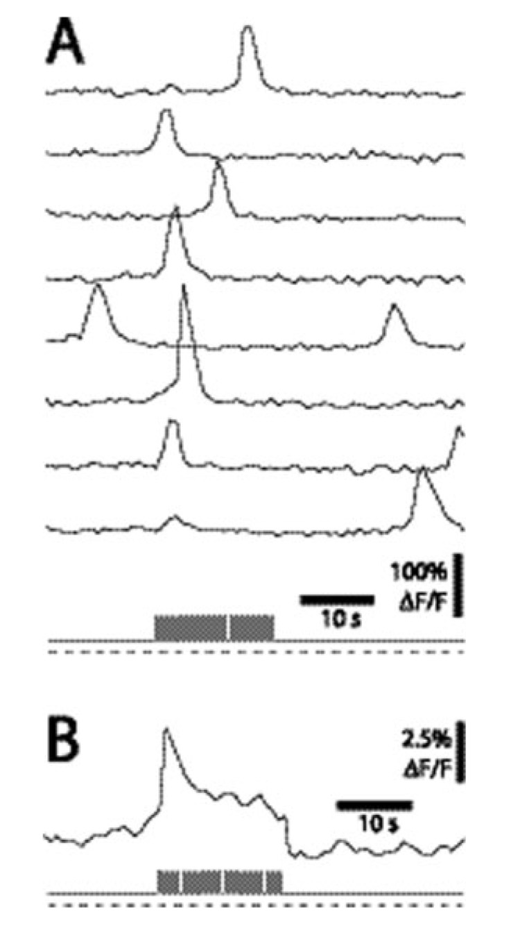
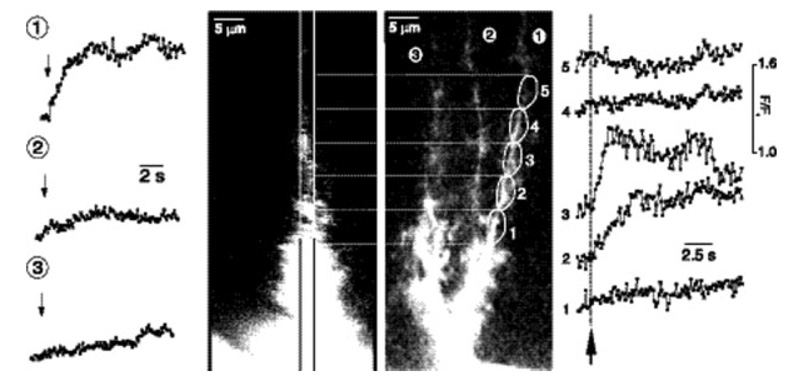
This article reviews calcium signaling in three specialized types of glial cells: Müller cells of the retina, Bergmann glial cells of the cerebellum, and radial glial cells of the developing cortex. Müller cells generate spontaneous and neuronal activity-evoked increases in Ca(2+). Neuron to Müller cell signaling is mediated by neuronal release of ATP and activation of glial P2Y receptors. Müller cells, in turn, modulate neuronal excitability and mediate vasomotor responses. Bergmann glial cells also generate spontaneous and activity-evoked Ca(2+) increases. Neuron to Bergmann glia signaling is mediated by neuronal release of nitric oxide, noradrenaline, and glutamate. In Bergmann glia, Ca(2+) increases control the structural and functional interactions between these cells and Purkinje cell synapses. In the ventricular zone of the developing cortex, radial glial cells generate spontaneous Ca(2+) increases that propagate as Ca(2+) waves through clusters of neighboring glial cells. These Ca(2+) increases control cell proliferation and neurogenesis.
This article reviews calcium signaling in three specialized types of glial cells: Müller cells of the retina, Bergmann glial cells of the cerebellum, and radial glial cells of the developing cortex. Müller cells generate spontaneous and neuronal activity-evoked increases in Ca2+ . Neuron to Müller cell signaling is mediated by neuronal release of ATP and activation of glial P2Y receptors. Müller cells, in turn, modulate neuronal excitability and mediate vasomotor responses. Bergmann glial cells also generate spontaneous and activity-evoked Ca2+ increases. Neuron to Bergmann glia signaling is mediated by neuronal release of nitric oxide, noradrenaline, and glutamate. In Bergmann glia, Ca2+ increases control the structural and functional interactions between these cells and Purkinje cell synapses. In the ventricular zone of the developing cortex, radial glial cells generate spontaneous Ca2+ increases that propagate as Ca2+ waves through clusters of neighboring glial cells. These Ca2+ increases control cell proliferation and neurogenesis.
Figures

References
-
- Anthony TE, Klein C, Fishell G, Heintz N. Radial glia serve asneuronal progenitors in all regions of the central nervous system. Neuron. 2004;41:881–890. - PubMed
-
- Bellamy TC, Ogden D. Short-term plasticity of Bergmann glial cell extrasynaptic currents during parallel fiber stimulation in rat cerebellum. Glia. 2005;52:325–335. - PubMed
-
- Bringmann A, Pannicke T, Weick M, Beidermann B, Uhlmann S, Kohen L, Wiedermann P, Reichenbach A. Activation of P2Y receptors stimulates potassium and cation currents in acutely isolated human Müller (glial) cells. Glia. 2002;37:139–152. - PubMed
-
- Burnashev N, Khodorova A, Jonas P, Helm PJ, Wisden W, Monyer H, Seeberg PH, Sakmann B. Calcium-permeable AMPA-kainate receptors in fusiform cerebellar glial cells. Science. 1992;256:1566–1570. - PubMed
Publication types
MeSH terms
Substances
Grants and funding
LinkOut - more resources
Full Text Sources
Miscellaneous
