Review
. 2006 Nov 15;54(7):747-757.
doi: 10.1002/glia.20423.

Anion channels in astrocytes: biophysics, pharmacology, and function

Affiliations
Review

Anion channels in astrocytes: biophysics, pharmacology, and function

Harold K Kimelberg et al. Glia. .

Abstract

The chloride/anion channels that have been so far identified in cultured astrocytes and those that have been confirmed in situ by a combination of mRNA identification, immunocytochemistry, and biophysical studies are reviewed. It is emphasized that we are just beginning to describe such channels and analyze their functions in astrocytes. The best-studied anion channels studied so far are those known as volume-regulated anion channels (VRACs). These, as for most channels, have been mainly studied in cultured astrocytes, but some correlative studies have been done in situ, because these channels have been emphasized as release routes for transmitters; namely, excitatory amino acids and ATP. They are activated by cell shape changes and cell swelling, and the release of amino acids and ATP and chloride currents, measured by whole cell clamping, by these processes has been well described, as is also their activation by low concentrations of extracellular ATP. However, the identity of these channels in astrocytes, as in all other cells, remains elusive. The potential involvement of VRACs in pathological states such as stroke, metastasis, and spreading depression is also discussed.


Figures

Fig. 1
Fig. 1
Cultured cortical rat astrocytes express ClC channels. A,B. Immunostaining with specific antibodies to ClC-2 and ClC-3 show prominent, membrane-associated staining. C. Transcripts for these channels are also found by RTPCR. Arrow indicates 500-bp band. D. Representative whole-cell recording of voltage steps from -80 to +80 mV for 200 ms from a 0-mV holding potential (inset) were obtained in choline—chloride bath and pipette solution following a 2-min hypotonic challenge. Currents were inhibited by substitution of 140 mM choline I for choline Cl-. (Adapted from Parkerson and Sontheimer, 2004.)
Fig. 2
Fig. 2
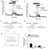
VRAC blockers DCPIB and tamoxifen potently inhibit swelling-activated D-[3H]aspartate release and Cl- currents in primary astrocyte cultures. A. Effect of 10 μM tamoxifen on swelling-activated D-[3H]aspartate release in the presence or absence of 10 μM ATP. Cells were exposed to hypo-osmotic medium (30% reduction in osmolarity for all experiments) as indicated, in the presence (□, ○) or absence (■, •) of 10 μM tamoxifen, applied as indicated. ATP was present in hypo-osmotic ∼ medium only (○, •). Data are means ±SEM of 5–7 experiments. **P = 0.002, tamoxifen vs. control; ###P < 0.001, ATP vs. ATP plus tamoxifen, repeated measures ANOVA. B. Effect of 10 μM tamoxifen on swelling-activated Cl- currents. Cells were held at 0 mV and step pulses to + and -40 mV were applied. Representative of six electrophysiological recordings. C. Effect of 20 μM DCPIB on swelling-activated D-[3H] aspartate release in the presence or absence of 20 μM ATP, in the presence (□, ○)or absence (■, •) of DCPIB, which was given 10 min before and during application of hypo-osmotic medium, as indicated. Data are means ± SEM of five experiments. ***P < 0.001, DCPIB vs. control; ***P < 0.001; ATP vs. ATP plus DCPIB, repeated measures ANOVA. D. Effect of 20-μM DCPIB on swelling-activated Cl- currents. Representative of six electrophysiological recordings. E. shows Cl- current responses to step pulses from -100 to +100 mV in 20-mV increments from 0-mV holding potential after exposure to hypotonic medium. F. Normalized release values relative to 100 for control swelling-activated excitatory amino acid release, with and without ATP and normalized VRAC currents in the absence of ATP in presence of tamoxifen or DCPIB. For B, D-F the isoosmotic external solution contained (in mM): 110 CsCl, 2 CaCl2, 1 MgSO4, 5 glucose, 10 Hepes, and 60 mannitol (pH 7.4, 290 mosmol). The hypoosmotic solution was made by omitting mannitol from isotonic solution and had an osmolarity of 230 mosmol. The pipette solution contained (in mM): 110 CsCl, 1 MgSO4, 1 Na2-ATP, 0.3 Na2-GTP, 15 Na-Hepes, 10 Hepes, and 1 EGTA (pH 7.3, 255 mosmol). The osmolarity of the pipette solution was set lower than that of the isotonic bath solution in order to prevent spontaneous cell swelling after attaining the whole-cell mode (from Abdullaev et al., 2006).
Fig. 3
Fig. 3
Activation of D-3H aspartate release by ATP with moderate and substantial swelling and effects of Ca2+ and changes in osmolarity. A. Moderate cell swelling was induced by 5% reduction in medium osmolarity (-14.5 mosmol). Ten micromoles of ATP was applied simultaneously with hypoosmotic medium. The data are the mean values ±SEM of seven experiments performed on three different cell culture preparations. B. Substantial cell swelling was induced by a 30% reduction in medium osmolarity (-90 mosmol). Ten micromolar ATP was applied simultaneously with hypoosmotic medium. The open squares show the ATP-induced D-[3H]aspartate release values in moderately swollen cells for comparison. The data are the mean values ±SEM of five experiments performed on two cell culture preparations. When not indicated, SEMs were less than symbols. C. Simultaneous measurements of the ATP-induced D-[3H]aspartate and [14C]taurine release from moderately swollen cultured astrocytes. Astrocytes were preloaded overnight with D-[3H]-aspartate and [14C]taurine. The data are means ±SEM of five experiments. D. ATP-induced organic osmolyte release in astrocytes is dependent on intracellular [Ca2+]. Cells were preincubated with 10 μM BAPTA-AM for 20 min, followed by a 5-min wash to remove extracellular BAPTA-AM. Then, astrocytes were exposed to a 5% reduction in medium osmolarity plus 10 μM ATP. The data are the means ±SEM of five experiments performed on two cell culture preparations. E. ATP-induced release is inhbited by an increase in medium osmolarity. Cells were exposed to 10 μM ATP and the simultaneous changes in medium osmolarity shown during 21st to 30th min of superfusion. F. Summary of the osmotic dependence of D-[3H]aspartate release in the presence or absence of 10 μM ATP shown in E. Open triangles represent an ATP-induced increment in EAA release. Data are the means ±SEM of 3–10 experiments performed on 2–4 different astrocyte culture preparations (from Mongin and Kimelberg, 2002, 2005).
Fig. 4
Fig. 4
Hyposmotic solutions cause swelling of astrocytes but not neurons. Two photon laser scanning microscopy of green fluorescent labeled (GFP) neurons and GFP astrocytes in cortical brain slices showed that osmotic changes in solution caused IOSs (intrinsic optical signals) that were not associated with any changes in neuronal volume but were associated with astrocytic swelling. The panels in column (A) show the changes in light transmittance as hippocampal brain slices swell or shrink in changing osmolarity. The GFP-labeled (B) soma, (C) dendrites, and (D) axon terminals of CA1 pyramidal neurons did not change their volume. In contrast (E) astrocytes showed swelling in hypo-osmotic solution and shrinkage in hyper-osmotic solution. The light transmittance and fluorescence images were obtained at 10 min after beginning perfusion with the solutions of different osmolarities, to ensure that a complete change had occurred in the slice (from Andrew et al., 2006).
Fig. 5
Fig. 5
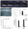
The release of glutamate during SD is reduced by NPPB, a blocker of volume-activated chloride channels. The release of glutamate during SD is reduced by NPPB, a blocker of volume-activated chloride channels, supporting a role for amino acid release due to astrocyte swelling. SD was triggered by inhibiting Na,K ATPase with ouabain. Imaging IOSs showed the progressive propagation of the depolarization and swelling during SD (A) (time in min; ΔT indicates increased light transmittance in arbitrary digital units) through the CA1 region (B) of the hippocampal brain slice. (C,D) SD still propagated in 0 external calcium, and NPPB in the absence of calcium reduced the onset slope of SD. The onset slope is the rate of change of transmittance during the propagation of SD. (E,F) NPPB also significantly reduced the efflux of glutamate during SD but did not alter GABA or glutamine efflux rates (from Basarsky et al., 1999). [Color figure can be viewed in the online issue, which is available at www.interscience.wiley.com.]

Similar articles

Cited by

References

    1. Abdullaev IF, Rudkouskaya A, Schools GP, Kimelberg HK, Mongin AA. Pharmacological comparison of swelling-activated excitatory amino acid release and Cl- currents in rat cultured astrocytes. J Physiol. 2006;572(Part 3):677–689. - PMC - PubMed
    1. Agnel M, Vermat T, Culouscou JM. Identification of three novel members of the calcium-dependent chloride channel (CaCC) family predominantly expressed in the digestive tract and trachea. FEBS Lett. 1999;455:295–301. - PubMed
    1. Ahmed N, Ramjeesingh M, Wong S, Varga A, Garami E, Bear CE. Chloride channel activity of ClC-2 is modified by the actin cytoskeleton. Biochem J. 2000;352(Part 3):789–794. - PMC - PubMed
    1. Aitken PG, Borgdorff AJ, Juta AJA, Kiehart DP, Somjen GG, Wadman WJ. Volume changes induced by osmotic stress in freshly isolated rat hippocampal neurons. Pflugers Arch. 1998;436:991–998. - PubMed
    1. Andersen P, Dingledine R, Gjerstad L, Langmoen IA, Laursen AM. Two different responses of hippocampal pyramidal cells to application of γ-amino butyric acid. J Physiol. 1980;305:279–296. - PMC - PubMed

Publication types

MeSH terms