

Human hepatic sinusoidal endothelial cells can be distinguished by expression of phenotypic markers related to their specialised functions in vivo
- PMID: 17006978
- PMCID: PMC4088223
- DOI: 10.3748/wjg.v12.i34.5429
Human hepatic sinusoidal endothelial cells can be distinguished by expression of phenotypic markers related to their specialised functions in vivo
Abstract
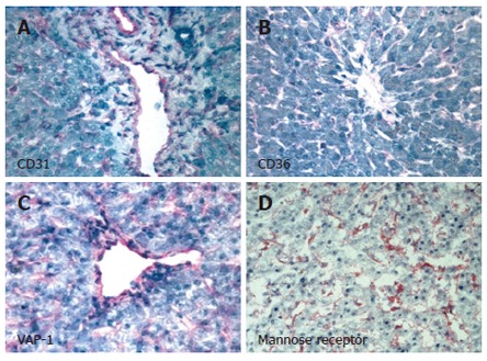
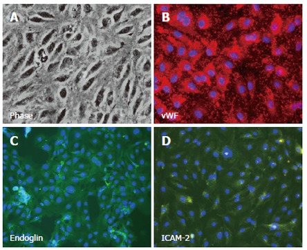
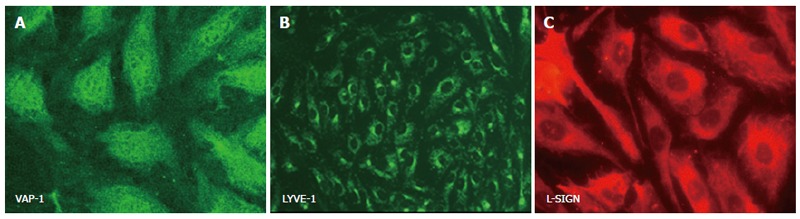
The hepatic sinusoids are lined by a unique population of hepatic sinusoidal endothelial cells (HSEC), which is one of the first hepatic cell populations to come into contact with blood components. However, HSEC are not simply barrier cells that restrict the access of blood-borne compounds to the parenchyma. They are functionally specialised endothelial cells that have complex roles, including not only receptor-mediated clearance of endotoxin, bacteria and other compounds, but also the regulation of inflammation, leukocyte recruitment and host immune responses to pathogens. Thus understanding the differentiation and function of HSEC is critical for the elucidation of liver biology and pathophysiology. This article reviews methods for isolating and studying human hepatic endothelial cell populations using in vitro models. We also discuss the expression and functions of phenotypic markers, such as the presence of fenestrations and expression of VAP-1, Stabilin-1, L-SIGN, which can be used to identify sinusoidal endothelium and to permit discrimination from vascular and lymphatic endothelial cells.
Figures
Similar articles
-
Vascular adhesion protein-1 mediates adhesion and transmigration of lymphocytes on human hepatic endothelial cells.J Immunol. 2002 Jul 15;169(2):983-92. doi: 10.4049/jimmunol.169.2.983. J Immunol. 2002. PMID: 12097405
-
Common lymphatic endothelial and vascular endothelial receptor-1 mediates the transmigration of regulatory T cells across human hepatic sinusoidal endothelium.J Immunol. 2011 Apr 1;186(7):4147-55. doi: 10.4049/jimmunol.1002961. Epub 2011 Mar 2. J Immunol. 2011. PMID: 21368224 Free PMC article.
-
Activation of vascular adhesion protein-1 on liver endothelium results in an NF-kappaB-dependent increase in lymphocyte adhesion.Hepatology. 2007 Feb;45(2):465-74. doi: 10.1002/hep.21497. Hepatology. 2007. PMID: 17256751
-
Cooperation of liver cells in health and disease.Adv Anat Embryol Cell Biol. 2001;161:III-XIII, 1-151. doi: 10.1007/978-3-642-56553-3. Adv Anat Embryol Cell Biol. 2001. PMID: 11729749 Review.
-
DC-SIGN, DC-SIGNR and LSECtin: C-type lectins for infection.Int Rev Immunol. 2014 Jan;33(1):54-66. doi: 10.3109/08830185.2013.834897. Epub 2013 Oct 24. Int Rev Immunol. 2014. PMID: 24156700 Review.
Cited by
-
Human parenchymal and non-parenchymal liver cell isolation, culture and characterization.Hepatol Int. 2013 Oct;7(4):951-8. doi: 10.1007/s12072-013-9475-7. Epub 2013 Oct 10. Hepatol Int. 2013. PMID: 26202025
-
Resolving the graft ischemia-reperfusion injury during liver transplantation at the single cell resolution.Cell Death Dis. 2021 Jun 8;12(6):589. doi: 10.1038/s41419-021-03878-3. Cell Death Dis. 2021. PMID: 34103479 Free PMC article.
-
Featured Article: Isolation, characterization, and cultivation of human hepatocytes and non-parenchymal liver cells.Exp Biol Med (Maywood). 2015 May;240(5):645-56. doi: 10.1177/1535370214558025. Epub 2014 Nov 12. Exp Biol Med (Maywood). 2015. PMID: 25394621 Free PMC article.
-
Sinusoidal endothelial cell repopulation following ischemia/reperfusion injury in rat liver transplantation.Hepatology. 2007 Nov;46(5):1464-75. doi: 10.1002/hep.21887. Hepatology. 2007. PMID: 17929236 Free PMC article.
-
Interactions of LSECtin and DC-SIGN/DC-SIGNR with viral ligands: Differential pH dependence, internalization and virion binding.Virology. 2008 Mar 30;373(1):189-201. doi: 10.1016/j.virol.2007.11.001. Epub 2008 Feb 20. Virology. 2008. PMID: 18083206 Free PMC article.
References
-
- Matsumoto T, Kawakami M. The unit-concept of hepatic parenchyma--a re-examination based on angioarchitectural studies. Acta Pathol Jpn. 1982;32 Suppl 2:285–314. - PubMed
-
- Takasaki S, Hano H. Three-dimensional observations of the human hepatic artery (Arterial system in the liver) J Hepatol. 2001;34:455–466. - PubMed
-
- Do H, Healey JF, Waller EK, Lollar P. Expression of factor VIII by murine liver sinusoidal endothelial cells. J Biol Chem. 1999;274:19587–19592. - PubMed
-
- Vidal-Vanaclocha F, Rocha M, Asumendi A, Barberá-Guillem E. Isolation and enrichment of two sublobular compartment-specific endothelial cell subpopulations from liver sinusoids. Hepatology. 1993;18:328–339. - PubMed
Publication types
MeSH terms
Substances
Grants and funding
LinkOut - more resources
Full Text Sources
Other Literature Sources
