

Localization of ANP-synthesizing cells in rat stomach
- PMID: 17007021
- PMCID: PMC4088169
- DOI: 10.3748/wjg.v12.i35.5674
Localization of ANP-synthesizing cells in rat stomach
Abstract
Aim: To study the morphological positive expression of antrial natriuretic peptide (ANP)-synthesizing cells and ultrastructural localization and the relationship between ANP-synthesizing cells and microvessel density in the stomach of rats and to analyze the distribution of the three histologically distinct regions of ANP-synthesizing cells.
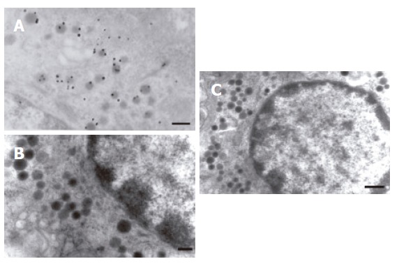
Methods: Using immunohistochemical techniques, we studied positive expression of ANP-synthesizing cells in rat stomach. A postembedding immunogold microscopy technique was used for ultrastructural localization of ANP-synthesizing cells. Microvessel density in the rat stomach was estimated using tannic acid-ferric chloride (TAFC) method staining. Distribution of ANP-synthesizing cells were studied in different regions of rat stomach histochemically.
Results: Positive expression of ANP-synthesizing cells were localized in the gastric mucosa of rats. Localization of ANP-synthesizing cells identified them to be enterochrochromaffin cells (EC) by using a postembedding immunogold electron microscopy technique. EC cells were in the basal third of the cardiac mucosa region. ANP-synthesizing cells existed in different regions of rat stomach and its density was largest in the gastric cardiac region, and the distribution order of ANP-synthesizing cells in density was cardiac region, pyloric region and fundic region in mucosa layer. We have also found a close relationship between ANP-synthesizing cells and microvessel density in gastric mucosa of rats using TAFC staining.
Conclusion: ANP-synthesizing cells are expressed in the gastric mucosa. EC synthesize ANP. There is a close relationship between ANP-synthesizing cells and microvessel density in gastric mucosa of rats. The distribution density of ANP-synthesizing cells is largest in the gastric cardiac region.
Figures


References
-
- McGrath MF, de Bold ML, de Bold AJ. The endocrine function of the heart. Trends Endocrinol Metab. 2005;16:469–477. - PubMed
-
- Bensimon M, Chang AI, de Bold ML, Ponce A, Carreras D, De Bold AJ. Participation of G proteins in natriuretic peptide hormone secretion from heart atria. Endocrinology. 2004;145:5313–5321. - PubMed
-
- Lai P, Nazian SJ, Gower WR Jr, Landon CS, Dietz JR. Increased bioactivity of rat atrial extracts: relation to aging and blood pressure regulation. J Gerontol A Biol Sci Med Sci. 2000;55:B390–B395. - PubMed
-
- Lai FJ, Hsieh MC, Hsin SC, Lin SR, Guh JY, Chen HC, Shin SJ. The cellular localization of increased atrial natriuretic peptide mRNA and immunoreactivity in diabetic rat kidneys. J Histochem Cytochem. 2002;50:1501–1508. - PubMed
-
- Cayli S, Ustünel I, Celik-Ozenci C, Korgun ET, Demir R. Distribution patterns of PCNA and ANP in perinatal stages of the developing rat heart. Acta Histochem. 2002;104:271–277. - PubMed
MeSH terms
Substances
LinkOut - more resources
Full Text Sources
