

Doppler study of hepatic vein in cirrhotic patients: correlation with liver dysfunction and hepatic hemodynamics
- PMID: 17007052
- PMCID: PMC4100667
- DOI: 10.3748/wjg.v12.i36.5853
Doppler study of hepatic vein in cirrhotic patients: correlation with liver dysfunction and hepatic hemodynamics
Abstract
Aim: To elucidate the significance of Doppler measurements of hepatic vein in cirrhotic patients and to correlate with liver dysfunction and hepatic hemodynamics.
Methods: One hundred patients with liver cirrhosis and 60 non-cirrhotic controls were studied. Doppler waveforms were obtained from right hepatic vein and flow velocity measured during quiet respiration. Doppler measurements were also obtained from portal trunk, right portal vein and proper hepatic artery.
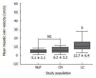
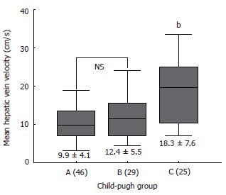
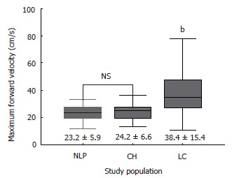
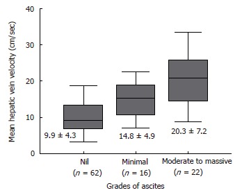
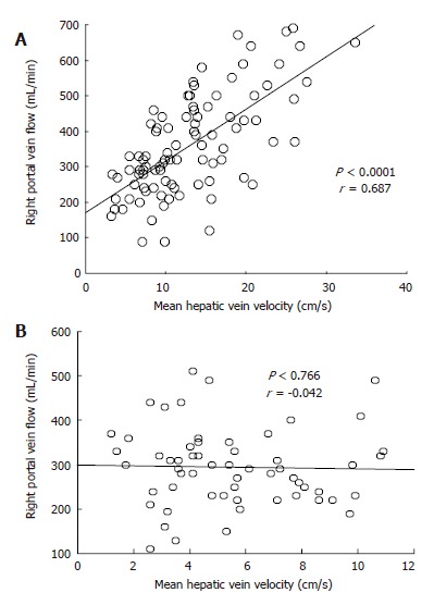
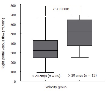
Results: Hepatic vein waveforms were classified into three classical patterns. Flat waveform was uncommon. Mean hepatic vein velocity was significantly higher in cirrhotic patients (12.7 +/- 6.4 vs 5.1 +/- 2.1 and 6.2 +/- 3.2 cm/s; P<0.0001). The poorer the grade of cirrhosis, the higher was the mean velocity. Maximum forward velocity was never greater than 40 cm/s in controls. Degree of ascites was found to be highly correlated with mean velocity. "Very highq group (>=20 cm/s) presented clinically with moderate to massive ascites. Correlations between right portal flow and mean velocity was significant (P<0.0001, r = 0.687).
Conclusion: Doppler waveforms of hepatic vein, which is independent of liver dysfunction, should be obtained during normal respiration. Mean hepatic vein velocity reflects the change in hepatic circulation associated with progression of liver cirrhosis. It can be used as a new parameter in the assessment of liver cirrhosis.
Figures
Similar articles
-
Prognostic significance of hepatic vein waveform by Doppler ultrasonography in cirrhotic patients with portal hypertension.Am J Gastroenterol. 1995 Oct;90(10):1853-7. Am J Gastroenterol. 1995. PMID: 7572908
-
Abnormality of the hepatic vein waveforms in cirrhotic patients with portal hypertension and its prognostic implications.J Gastroenterol Hepatol. 2008 Jul;23(7 Pt 2):e129-36. doi: 10.1111/j.1440-1746.2007.05155.x. Epub 2007 Oct 9. J Gastroenterol Hepatol. 2008. PMID: 17924952
-
Qualitative hepatic venous Doppler sonography versus portal flowmetry in predicting the severity of esophageal varices in hepatitis C cirrhosis.AJR Am J Roentgenol. 1997 Aug;169(2):511-5. doi: 10.2214/ajr.169.2.9242766. AJR Am J Roentgenol. 1997. PMID: 9242766
-
The value of Doppler ultrasound in cirrhosis and portal hypertension.Scand J Gastroenterol Suppl. 1999;230:82-8. doi: 10.1080/003655299750025598. Scand J Gastroenterol Suppl. 1999. PMID: 10499467 Review.
-
Altered Doppler flow patterns in cirrhosis patients: an overview.Ultrasonography. 2016 Jan;35(1):3-12. doi: 10.14366/usg.15020. Epub 2015 May 27. Ultrasonography. 2016. PMID: 26169079 Free PMC article. Review.
Cited by
-
Correlation Between Hepatic Waveform Changes on Doppler Ultrasound and Disease Severity in Cirrhotic Patients.Cureus. 2025 May 7;17(5):e83658. doi: 10.7759/cureus.83658. eCollection 2025 May. Cureus. 2025. PMID: 40486465 Free PMC article.
-
Hepatic perfusion as a new predictor of prognosis and mortality in critical care patients with acute-on-chronic liver failure.Front Med (Lausanne). 2022 Oct 10;9:1008450. doi: 10.3389/fmed.2022.1008450. eCollection 2022. Front Med (Lausanne). 2022. PMID: 36300192 Free PMC article.
-
[Ultrasound diagnostics of the liver : Principles and important pathologies].Radiologie (Heidelb). 2023 May;63(5):387-402. doi: 10.1007/s00117-023-01138-3. Epub 2023 Apr 18. Radiologie (Heidelb). 2023. PMID: 37071126 German.
-
Diagnostic accuracy of the attenuation value in abdominal contrast enhanced dynamic multi-detector-row computed tomography for esophageal varices in patients with liver cirrhosis.Eur J Radiol Open. 2021 Apr 23;8:100347. doi: 10.1016/j.ejro.2021.100347. eCollection 2021. Eur J Radiol Open. 2021. PMID: 33997144 Free PMC article.
-
Pre-operative Hepatic Artery Resistive Index is a Non-invasive Predictive Indicator of Prognosis in Biliary Atresia.J Indian Assoc Pediatr Surg. 2017 Oct-Dec;22(4):237-241. doi: 10.4103/jiaps.JIAPS_103_17. J Indian Assoc Pediatr Surg. 2017. PMID: 28974877 Free PMC article.
References
-
- Burns PN. Hemodynamics. In: Taylor KJW, Burns PN, Wells PNT, editors. Clinical applications of Doppler ultrasound. New York: Raven Press; 1988. pp. 46–75.
-
- Bolondi L, Li Bassi S, Gaiani S, Zironi G, Benzi G, Santi V, Barbara L. Liver cirrhosis: changes of Doppler waveform of hepatic veins. Radiology. 1991;178:513–516. - PubMed
-
- Ohta M, Hashizume M, Tomikawa M, Ueno K, Tanoue K, Sugimachi K. Analysis of hepatic vein waveform by Doppler ultrasonography in 100 patients with portal hypertension. Am J Gastroenterol. 1994;89:170–175. - PubMed
-
- von Herbay A, Frieling T, Häussinger D. Association between duplex Doppler sonographic flow pattern in right hepatic vein and various liver diseases. J Clin Ultrasound. 2001;29:25–30. - PubMed
-
- Colli A, Cocciolo M, Riva C, Martinez E, Prisco A, Pirola M, Bratina G. Abnormalities of Doppler waveform of the hepatic veins in patients with chronic liver disease: correlation with histologic findings. AJR Am J Roentgenol. 1994;162:833–837. - PubMed
MeSH terms
LinkOut - more resources
Full Text Sources
Medical
