. 2006 Oct 1;78(19):6826-32.
doi: 10.1021/ac060652r.

Massively parallel sample preparation for the MALDI MS analyses of tissues

Affiliations

Massively parallel sample preparation for the MALDI MS analyses of tissues

Eric B Monroe et al. Anal Chem. .

Abstract

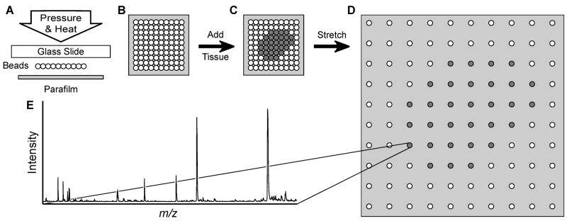
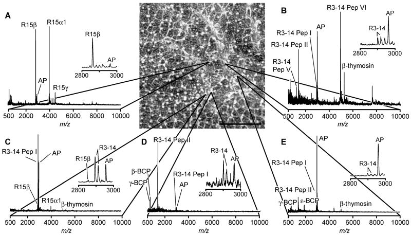
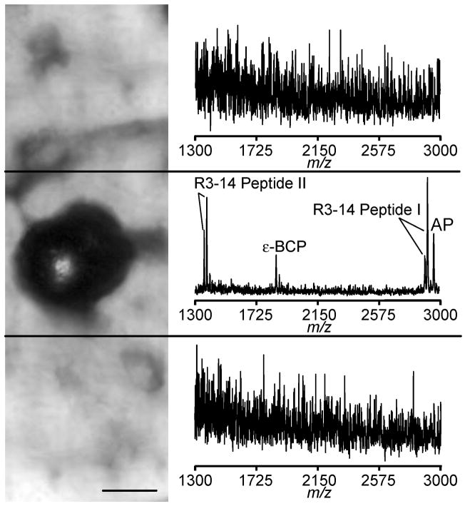
Investigation of the peptidome of the nervous system containing large, often easily identifiable neurons has greatly benefited from single-cell matrix-assisted laser desorption/ionization (MALDI) mass spectrometry and has led to the discovery of hundreds of novel cell-to-cell signaling peptides. By combining new sample preparation methods and established protocols for bioanalytical mass spectrometry, a high-throughput, small-volume approach is created that allows the study of the peptidome of a variety of nervous systems. Specifically, approximately single-cell-sized samples are rapidly prepared from thin tissue slices by adhering the tissue section to a glass bead array that is anchored to a stretchable membrane. Stretching the membrane fragments the tissue slice into thousands of individual samples, their dimensions predominately governed by the size of the individual glass beads. Application of MALDI matrix, followed by the repeated condensation of liquid microdroplets on the fragmented tissue, allows for maximal analyte extraction and incorporation into MALDI matrix crystals. During extraction, analyte migration between the pieces of tissue on separate beads is prevented by the underlying hydrophobic substrate and by controlling the size of the condensation droplets. The procedure, while general in nature, may be tailored to the needs of a variety of analyses, producing mass spectra equivalent to those acquired from single-cell samples.


Figures

Figure 1
Figure 1
Schematic of the massively parallel sample preparation using the stretched sample method. (A, B) Pressure and heat are used to form a layer of glass beads on a Parafilm M membrane surface. (C) A thin tissue slice is placed onto the glass bead layer. (D) The Parafilm M membrane is manually stretched. As a result, the tissue slice is fragmented into thousands of spatially isolated pieces. (E) After MALDI matrix application, individual pieces of tissue may then be investigated with MALDI MS.
Figure 2
Figure 2
Tissue sectioning, deposition on the substrate, and stretching. (A) Photograph of the internal space of the cryotome used for tissue sectioning. The frozen abdominal ganglion is positioned on a piece of buccal muscle attached to a metal stage, which is visible in the upper left corner. A prepared substrate and deposited section of abdominal ganglion is located centrally. (B) Microphotograph of an abdominal ganglion section deposited on a glass slide. Cytoplasm and nuclear regions of neurons are visible. (C) Microphotograph of an abdominal ganglion section deposited onto glass bead layer (orange region in the center of view). (D) Following stretching and matrix application, the tissue is largely fragmented into individual bead-sized pieces.
Figure 3
Figure 3
MALDI matrix application/analyte extraction procedures. (A) Microphotographs of a glass bead layer after Parafilm M stretching and DHB application. (B) In order to increase sensitivity and promote analyte extraction from the tissue, the sample is cooled in a humid atmosphere to form small droplets on the glass bead surface. (C) The sample is then allowed to dry over a period of several minutes. (D) Time course of sample temperature (bottom, black line) and humidity (upper, gray line) changes within the sample chamber. (Scalebars = 100 μm.)
Figure 4
Figure 4
Analyte migration between individual glass beads is restricted by Parafilm M hydrophobic surface. (A) Ion image of background signal (m/z 1204) shows a largely uniform signal intensity distribution across the patterned sample while (B) angiotensin I (m/z 1296) is confined to the region of the stretched substrate which contains beads. (Scalebars = 1 mm.)
Figure 5
Figure 5
Representative mass spectra (A–E) from a 10-μm thick abdominal ganglion section, prepared using the stretched sample method, demonstrate a wide variation in the peptide content of neurons within a ~750 μm × 750 μm region of ganglion. Microphotograph of the section after stretching is shown in the top center part of this figure with mass spectra linked to the corresponding bead from which the signal arises. (Scalebar = 1 mm.)
Figure 6
Figure 6
A microphotograph showing three areas of the Parafilm M surface where mass spectra were acquired, with only the middle region having an individual glass bead with attached tissue piece; the top and bottom adjacent areas are devoid of the beads. On the right are the resulting mass spectra from each region; peptides are only detected in the region with the bead. (Scalebar = 25 μm.)

References

    1. van Veelen PA, Jimenez CR, Li KW, Wildering WC, Gerearts WP, Tjaden UR, van der Greef J. Organic Mass Spectrom. 1993;28:1542–1546.
    1. Jimenez CR, van Veelen PA, Li KW, Wildering WC, Gerearts WP, Tjaden UR, van der Greef J. J Neurochem. 1994;62:404–407. - PubMed
    1. Garden RW, Shippy SA, Li L, Moroz TP, Sweedler JV. Proc Natl Acad Sci USA. 1998;95:3972–3977. - PMC - PubMed
    1. van Strien FJ, Jespersen S, van der Greef J, Jenks BG, Roubos EW. FEBS Lett. 1996;379:165–170. - PubMed
    1. Sweedler JV, Rubakhin SS, Churchill JD, Greenough WT. Program No. 326.162003. 2003 Abstract Viewer and Itinerary Planner. Washington, DC: Society for Neuroscience; 2003. Online.

Publication types

MeSH terms