. 2006 Oct 1;78(19):6948-54.
doi: 10.1021/ac0613131.

Capillary electrophoresis separation in the presence of an immiscible boundary for droplet analysis

Affiliations

Capillary electrophoresis separation in the presence of an immiscible boundary for droplet analysis

J Scott Edgar et al. Anal Chem. .

Abstract

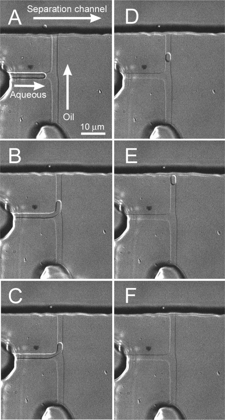
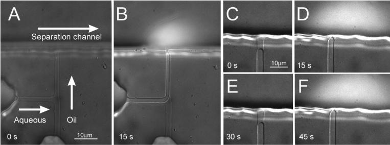
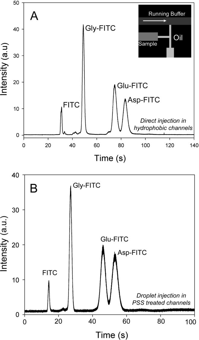
This paper demonstrates the ability to use capillary electrophoresis (CE) separation coupled with laser-induced fluorescence for analyzing the contents of single femtoliter-volume aqueous droplets. A single droplet was formed using a T-channel (3 microm wide by 3 microm tall) connected to microinjectors, and then the droplet was fluidically moved to an immiscible boundary that isolates the CE channel (50 microm wide by 50 microm tall) from the droplet generation region. Fusion of the aqueous droplet with the immiscible boundary effectively injects the droplet content into the separation channel. In addition to injecting the contents of droplets, we found aqueous samples can be introduced directly into the separation channel by reversibly penetrating and resealing the immiscible partition. Because droplet generation in channels requires hydrophobic surfaces, we have also investigated the advantages to using all hydrophobic channels versus channel systems with patterned hydrophobic and hydrophilic regions. To fabricate devices with patterned surface chemistry, we have developed a simple strategy based on differential wetting to deposit selectively a hydrophilic polymer (poly(styrenesulfonate)) onto desired regions of the microfluidic chip. Finally, we applied our device to the separation of a simple mixture of fluorescein-labeled amino acids contained within a approximately 10-fL droplet.


Figures

Figure 1
Figure 1. Schematic showing the design of our microfluidic chip and an overview of our experimental setup
(A) The separation channel was 50 μm by 50 μm in cross section; the small T-channel where the sample and oil met had a height of 3 μm and a width of 3 μm. The inset highlights the difference in dimension between these two channels. (B) Our experiments were performed on an inverted microscope using an Ar+ laser for exciting fluorescence; a CCD camera was used for wide-field imaging and an avalanche photodiode (APD) for point detection. A set of home-built microinjectors and a high-voltage power supply were connected to the microfluidic chip for droplet generation and capillary-electrophoresis separation.
Figure 2
Figure 2
A sequence of images showing the generation and transport of a single aqueous droplet to the separation channel. Pressure was first applied to the sample-containing aqueous channel, which caused the aqueous interface to protrude into the channel that contained the continuous phase (A, B). Subsequent flow of oil then sheared off an aqueous droplet (C) and moved the droplet to the immiscible boundary (D, E), where it was injected into the separation channel (F) before a high-voltage was applied to separate the contents of the droplet.
Figure 3
Figure 3. Direct injection of aqueous sample into the separation channel
(A) In direct injection, the T-channel was first filled with oil. (B) Pressure applied to the aqueous sample caused displacement of the oil, and brought the sample into direct contact with the separation buffer. (C-F) shows two cycles of direction injection. Note any retraction of the sample solution led to re-sealing of the immiscible interface and re-establishment of the immiscible boundary (C, E). The sample solution contained fluorescein-labeled amino acids and was visualized under bright-field and epi illumination. Plumes of injected sample are seen in B, D, and F.
Figure 4
Figure 4. Electropherogram showing the separation of fluorescein isothiocyanate (FITC) and FITC-labeled glycine, glutamate, and aspartate
(A) Separation was performed in a hydrophobic PDMS separation channel after direct injection; the applied field strength was 650 V/cm and the separation distance was 3.2 cm. During separation, the sample-oil interface was retreated back to the sample channel (inset). (B) Separation of the contents of a single 10 fL volume droplet, which was performed in a polystyrene sulfonate coated separation channel; the applied voltage was 500 V/cm and the separation distance was 2 cm. The sample-oil interface has retracted during the separation as in (A). The Y-axis in (A) and (B) have different scales.
Figure 5
Figure 5. Selective patterning of microchannels and the formation of a stable immiscible boundary
(A) Schematic showing the selective wetting of the large separation channel by a polyelectrolyte-containing aqueous solution; all channel surfaces initially were hydrophobic. (B, C) Under positive pressure from the oil phase, the polystyrene sulfonate patterned channel exhibited a high degree of curvature (B), but in untreated all-hydrophobic channels, the oil wetted the walls of the separation channel that resulted in a low degree of curvature (C). (D-F) A sequence of images that show the selective patterning of the separation channel. The channels initially were all empty (in contact with air) and hydrophobic (D). A solution containing fluorescently-labeled IgG antibodies were then flowed through the separation channel; note the formation of the stable air-water interface (arrow) at the entrance to the T-channel (E). The fluorescent antibodies were laid down only on the surfaces in the separation channel, as visualized by epi-fluorescence after the solution has been removed (F).

References

    1. Lorenz RM, Edgar JS, Jeffries GD, Chiu DT. Anal. Chem. 2006 In press. - PubMed
    1. Chiu DT. TrAC. 2003;22:528–536.
    1. He M, Edgar JS, Jeffries GD, Lorenz RM, Shelby JP, Chiu DT. Anal Chem. 2005;77:1539–1544. - PubMed
    1. Zheng B, Roach LS, Ismagilov RF. J. Am. Chem. Soc. 2003;125:11170–11171. - PubMed
    1. Dittrich PS, Jahnz M, Schwille P. Chembiochem. 2005;6:811–814. - PubMed

Publication types

MeSH terms