. 2006 Dec 15;400(3):563-72.
doi: 10.1042/BJ20061020.

The Dbs PH domain contributes independently to membrane targeting and regulation of guanine nucleotide-exchange activity

Affiliations

The Dbs PH domain contributes independently to membrane targeting and regulation of guanine nucleotide-exchange activity

Mark A Baumeister et al. Biochem J. .

Abstract

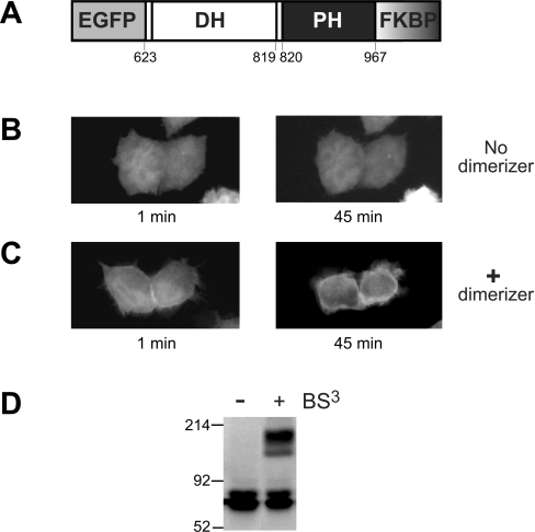
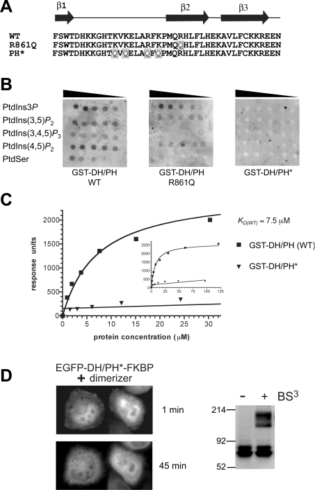
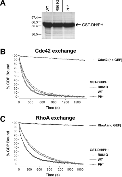
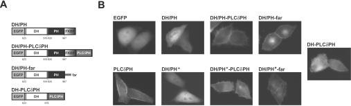
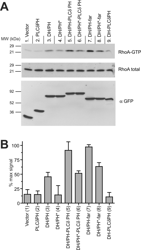
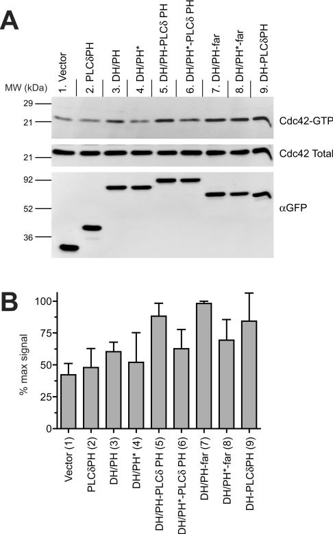
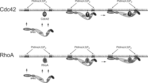
Dbl family GEFs (guanine nucleotide-exchange factors) for the Rho GTPases almost invariably contain a PH (pleckstrin homology) domain adjacent to their DH (Dbl homology) domain. The DH domain is responsible for GEF activity, and the PH domain plays a regulatory role that remains poorly understood. We demonstrated previously that Dbl family PH domains bind phosphoinositides with low affinity and cannot function as independent membrane targeting modules. In the present study, we show that dimerization of a Dbs (Dbl's big sister) DH/PH domain fragment is sufficient to drive it to the plasma membrane through a mechanism involving PH domain-phosphoinositide interactions. Thus, the Dbs PH domain could play a significant role in membrane targeting if it co-operates with other domains in the protein. We also show that mutations that prevent phosphoinositide binding by the Dbs PH domain significantly impair cellular GEF activity even in chimaeric proteins that are robustly membrane targeted by farnesylation or by the PH domain of phospholipase C-delta1. This finding argues that the Dbs PH domain plays a regulatory role that is independent of its ability to aid membrane targeting. Thus, we suggest that the PH domain plays dual roles, contributing independently to membrane localization of Dbs (as part of a multi-domain interaction) and allosteric regulation of the DH domain.


Figures

Figure 1
Figure 1. The DH/PH fragment of Dbs can be driven to the plasma membrane by dimerization
(A) FKBPF36V was fused to the C-terminus of an EGFP–Dbs DH/PH fragment to give the EGFP–DH/PH–FKBPF36V fusion protein schematized here. Amino acid numbers corresponding to domain boundaries are given. HeLa cells transiently transfected with the EGFP–DH/PH–FKBPF36V construct (containing the wild-type Dbs PH domain) were either untreated (B) or treated (C) with the AP20187 dimerizer at a final concentration of 200 nM (see Experimental section), and live cells were visualized by fluorescence microscopy after the indicated times. (D) AP20187 addition promotes dimerization of the EGFP–DH/PH–FKBPF36V fusion protein as assessed by cross-linking with BS3 (see Experimental section). Whole-cell lysates were subjected to treatment with BS3, and analysed by SDS/PAGE and immunoblotting with an anti-GFP antibody.
Figure 2
Figure 2. Mutations in the PH domain of Dbs abolish its weak PtdIns(4,5)P2 binding
(A) The sequence from strand β1 to β3 of the Dbs PH domain is shown, corresponding to the region of PH domains known to interact with phosphoinositides [32]. Mutations (to glutamine) were introduced into the β2 strand (R861Q) or at four basic residues (K849, K851, R855, and K857) in the β1/β2 loop (PH*) of the Dbs PH domain as indicated by the grey boxed Qs. (B) 32P-Labelled GST–DH/PH (10 μg) was used to probe nitrocellulose filters spotted with serial 2-fold dilutions of PtdIns3P, PtdIns(3,5)P2, PtdIns(3,4,5)P3, PtdIns(4,5)P2 and phosphatidylserine (PtdSer) as marked (beginning at 2 mg/ml). (C) GST–DH/PH and GST–DH/PH* proteins were assessed for binding to PtdIns(4,5)P2 using SPR as described [8]. GST–DH/PH or GST–DH/PH* at a series of concentrations were flowed over a Biacore sensor chip containing 3% (mol/mol) PtdIns(4,5)P2 in dioleoylphosphatidylcholine. Steady-state binding signals are plotted against protein concentration, with the best-fit to a simple 1:1 binding curve superimposed. The KD (app) value for binding of (dimeric) GST–DH/PH (wild-type) was approx. 7.5 μM. The inset curve demonstrates saturation of PtdIns(4,5)P2 binding by GST–DH/PH. GST–DH/PH* gave no significant binding signal at any concentration tested. Curves are representative of at least three independent experiments. (D) Introduction of PH* mutations (which prevent phosphoinositide binding as shown in B and C) abolished the ability of the EGFP–DH/PH*–FKBPF36V to translocate to the plasma membrane upon addition of AP20187. Details are as described for Figures 1(B) and 1(C). BS3 cross-linking experiments were performed as in Figure 1(D) and show that AP20187 induces robust EGFP–DH/PH*–FKBPF36V dimerization.
Figure 3
Figure 3. PH domain mutations do not impair solubility or in vitro GEF activity of the Dbs DH/PH fragment
(A) A Coomassie Blue-stained SDS/PAGE gel of E. coli-expressed wild-type (WT), R861Q and PH* forms of GST–DH/PH after one-step purification with glutathione–agarose beads is shown. Expression levels, solubility and stability appeared identical for the three proteins. In vitro GTP/GDP exchange on Cdc42 (B) and RhoA (C) was measured for wild-type (WT) and mutated GST–DH/PH proteins as described in the Experimental section. The data were fitted as single exponential decays, giving kobs for Cdc42 of 0.15×10⁻³ s−1 (no GEF), 4.29×10⁻³ s−1 (wild-type), 3.72×10⁻³ s−1 (R861Q) and 6.02×10⁻³ s−1 (PH*). Rates for RhoA were 0.31×10⁻³ s−1 (no GEF), 2.25×10⁻³ s−1 (wild type), 1.97×10⁻³ s−1 (R861Q) and 3.99×10⁻³ s−1 (PH*). Each experiment was performed twice with identical results.
Figure 4
Figure 4. Membrane targeting of the Dbs DH/PH fragment
(A) Schematic representation of the chimaeric constructs employed for membrane targeting of DH/PH and DH/PH*. To target the Dbs DH/PH fragment with PLC-δ1-PH, the PLC-δ1 PH domain was fused to the C-terminus of the EGFP–DH/PH–FKBPF36V chimaera shown in Figure 1(A), using the FKBP moiety as a spacer between the two PH domains. For targeting the DH/PH fragment by farnesylation (far), the farnesylation sequence of H-Ras was fused directly to the C-terminus of the EGFP–DH/PH fusion. To analyse the isolated DH domain, PLC-δ1-PH was fused directly to its C-terminus. Constructs including the Dbs PH domain were made with both the wild-type and PH* versions of the PH domain. The nomenclature used in the text for each construct is noted above its scheme. (B) Membrane localization of each EGFP chimaera was assessed by fluorescence microscopy of transiently-transfected live HeLa cells. No significant plasma membrane localization is seen for EGFP, or EGFP–DH/PH (wild-type or PH*). All fusion proteins containing PLC-δ1-PH or the far signal were robustly targeted to the plasma membrane. Structures resembling filopodia were seen in more than 60% of cells (n=150) exhibiting membrane localization of a DH-domain-containing fragment (DH/PH–PLC δ1 PH, DH/PH*–PLC δ1 PH, DH/PH–far, DH/PH*–far and DH–PLC δ1 PH).
Figure 5
Figure 5. RhoA activation by membrane-targeted Dbs DH/PH
(A) Representative immunoblots for RhoA activation experiments. HeLa cells were transfected with the indicated construct (see Figure 4A), and the active pool of (GTP-bound) RhoA was specifically isolated from serum-starved cells by affinity chromatography using GST–Rhotekin RBD as described in the Experimental section. The top and middle panels were immunoblotted with an antibody to RhoA, and represent (top) RhoA protein precipitated with GST–Rhotekin RBD (GTP-bound, active) and (middle) whole-cell lysate (total RhoA). The bottom panel represents whole-cell lysate immunoblotted with an antibody against GFP for comparison of expression levels of the different chimaeric Dbs DH/PH. (B) Immunoblots from at least three independent experiments were quantified using Kodak ImageStation software. The normalized intensity of each band was evaluated as a percentage of the maximum RhoA activation in that experiment, and results are presented as means±S.D.
Figure 6
Figure 6. Cdc42 activation by membrane-targeted Dbs DH/PH
(A) Representative immunoblots for Cdc42 activation experiments. Experiments were performed as for Figure 5(A), but using GST–PAK PBD to specifically isolate activated Cdc42. Top and middle panels were immunoblotted with an antibody to Cdc42, and represent activated (top) and total (middle) Cdc42 respectively. The bottom panel represents whole-cell lysate immunoblotted with an antibody against GFP for assessing Dbs fragment expression levels. (B) Quantification of Cdc42 activation experiment, as described in the legend to Figure 5(B).
Figure 7
Figure 7. Schematic model for the role of the Dbs PH domain in regulating GEF activity
Membrane recruitment of Dbs is promoted by the co-operation of multiple domains, including the Sec14 domain [27], possibly the SH3 domain, plus PH domain–phosphoinositide interactions, PH domain–GTPase (Cdc42 or RhoA) interactions, and DH domain–GTPase (Cdc42 or RhoA) interactions. At the plasma membrane, optimal binding to the GTPase target requires ligation of the PH domain by PtdIns(4,5)P2 in order to relieve possible steric hindrance to DH domain interactions and maximize interactions between the GTPase and the PH domain. In the case of Cdc42, relief of inhibition may play a more important role, as discussed in the text. Our results suggest that the PH domain interactions may be more important in the case of RhoA, as indicated in the Figure. D, GDP bound to Cdc42 or RhoA; T; GTP-bound state.

References

    1. Whitehead I. P., Campbell S., Rossman K. L., Der C. J. Dbl family proteins. Biochim. Biophys. Acta. 1997;1332:F1–F23. - PubMed
    1. Hall A. Rho GTPases and the actin cytoskeleton. Science. 1998;279:509–514. - PubMed
    1. Hart M. J., Eva A., Zangrilli D., Aaronson S. A., Evans T., Cerione R. A., Zheng Y. Cellular transformation and guanine nucleotide exchange activity are catalysed by a common domain on the dbl oncogene product. J. Biol. Chem. 1994;269:62–65. - PubMed
    1. Snyder J. T., Worthylake D. K., Rossman K. L., Betts L., Pruitt W. M., Siderovski D. P., Der C. J., Sondek J. Structural basis for the selective activation of Rho GTPases by Dbl exchange factors. Nat. Struct. Biol. 2002;9:468–475. - PubMed
    1. Rossman K. L., Sondek J. Larger than Dbl: new structural insights into RhoA activation. Trends Biochem. Sci. 2005;30:163–165. - PubMed

Publication types

Substances