. 2006 Nov 24;364(2):136-51.
doi: 10.1016/j.jmb.2006.08.084. Epub 2006 Sep 3.

Elucidation of human choline kinase crystal structures in complex with the products ADP or phosphocholine

Affiliations

Elucidation of human choline kinase crystal structures in complex with the products ADP or phosphocholine

Enrico Malito et al. J Mol Biol. .

Abstract

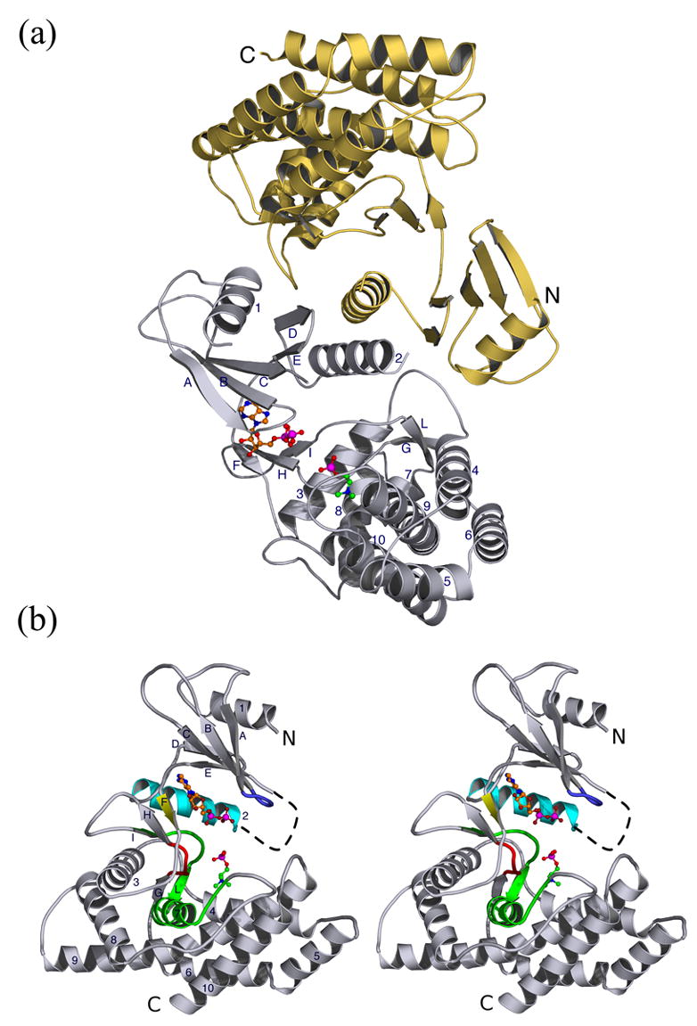
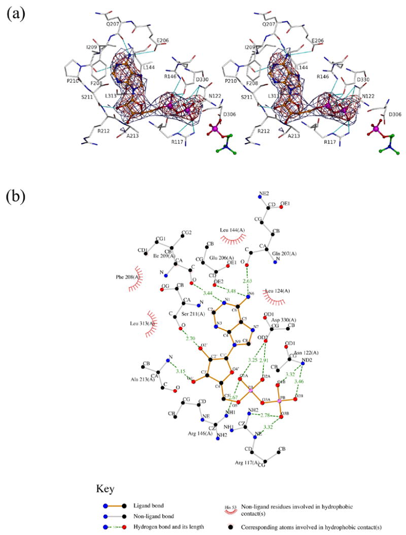
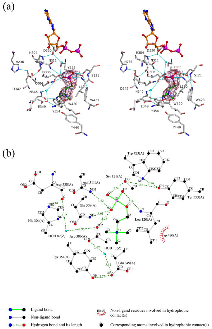
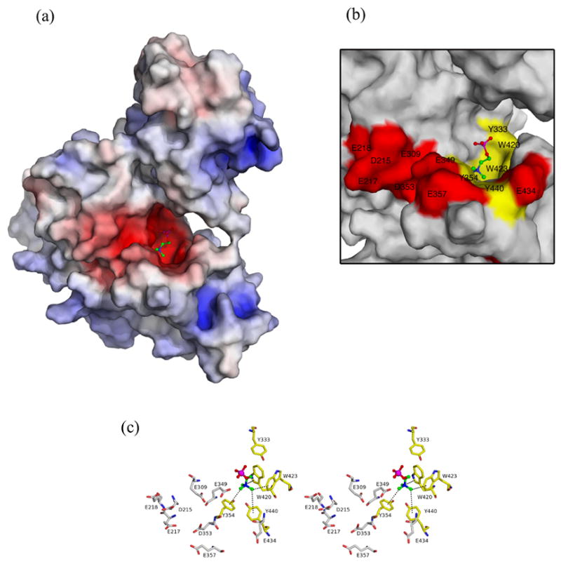
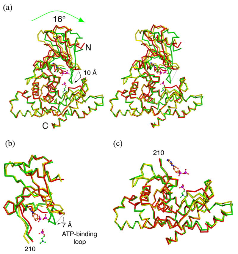
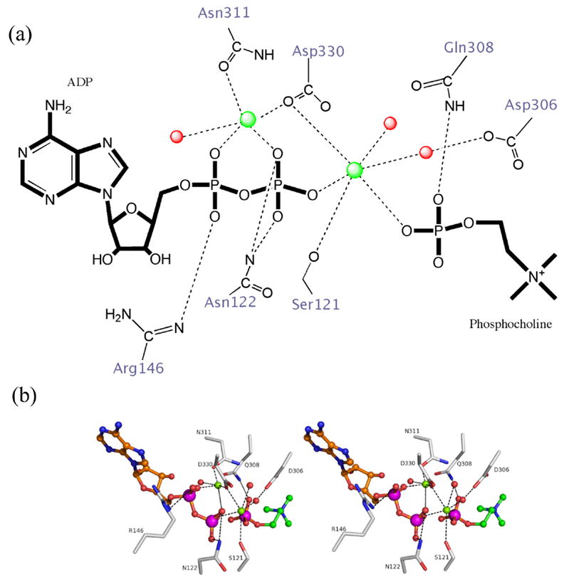
Choline kinase, responsible for the phosphorylation of choline to phosphocholine as the first step of the CDP-choline pathway for the biosynthesis of phosphatidylcholine, has been recognized as a new target for anticancer therapy. Crystal structures of human choline kinase in its apo, ADP and phosphocholine-bound complexes, respectively, reveal the molecular details of the substrate binding sites. ATP binds in a cavity where residues from both the N and C-terminal lobes contribute to form a cleft, while the choline-binding site constitutes a deep hydrophobic groove in the C-terminal domain with a rim composed of negatively charged residues. Upon binding of choline, the enzyme undergoes conformational changes independently affecting the N-terminal domain and the ATP-binding loop. From this structural analysis and comparison with other kinases, and from mutagenesis data on the homologous Caenorhabditis elegans choline kinase, a model of the ternary ADP.phosphocholine complex was built that reveals the molecular basis for the phosphoryl transfer activity of this enzyme.


Figures

Fig. 1
Fig. 1
Ribbon diagram of apo hCKα2, and with ADP and PChol molecules concomitantly present as modeled on the basis of the determined hCKADP and hCKPChol complex structures. (a) hCKApo dimer (gray/yellow ribbon) oriented with the two-fold non-crystallographic axes perpendicular to the plane of the page. Dimer formation buries 2100 Å, where helix 2 plays a major role. (b) Stereo diagram of a hCKα2 monomer drawn after a vertical rotation of 90° with respect to the orientation in (a). Key structural elements are colored: the ATP-binding loop in blue, the dimer interface α-helix in cyan, the short β-strand that links the N- and C-terminal domains in yellow, the Brenner’s motif in red, and the choline kinase motif in green. In this and the following figures, ball-and-stick representation of ADP and PChol molecules are shown with their carbon atoms colored in orange and green, respectively. Oxygen, nitrogen and phosphate atoms are shown in red, blue and magenta, respectively. All structure figures were made with Pymol.
Fig. 2
Fig. 2
Sequence alignment of choline kinases, APH(3′)-IIIa and cAPK obtained with the ClustalW algorithm. Key structural elements are shaded with different colors according to Figure 1(b). Residue that directly interact with ATP are marked by an “a”, those with choline by a “c”, and the hydrophobic residues that surround the choline by an “h”. Residues revealed by mutagenesis experiments to play a structural role are labeled with an “s”. hCK denotes human choline kinases, mCK from mouse, and cCK from C. Elegans. The sequence alignment for APH(3′)-IIIa (APH) and cAPK is shown only for catalytically important residues. Sequence breaks for the latter two are denoted with // and sequence not included in this alignment with period marks (..). Gaps in a sequence are marked with --. The region marked with black dashed line represents the 18 amino-acid insertion that distinguishes hCKα-2 from hCKα-1 and that is not visible in all our crystal structures. The first residue in our Δ49-hCKα-2 construct is indicated by an arrow, and so is the first visible residue in our crystal structures.
Fig. 3
Fig. 3
The ATP binding site. (a) Stereo view representation of the ATP-binding site. 2Fo-Fc simulated annealing omit map (red) and sigma-a weighted 2Fo-Fc map (blue) both contoured at 1 σ are shown around ADP. The relative position of PChol is shown for orientation purposes. The protein carbon, oxygen, nitrogen and phosphate atoms are shown in grey, red, blue and magenta, respectively. H-bonds are shown as cyan dashed lines. (b) Ligplot drawing showing the interactions of ADP with hCKα2. Dashed lines indicate all potential H-bonds. “Radiating” spheres indicate hydrophobic contacts between carbon atoms of the nucleotide and the neighboring residues. Ligand-bonds, carbon, oxygen, nitrogen and phosphorous atoms are colored orange, black, red, blue and purple, respectively. Note that we could not observe side chain density for Arg213 so this residue was modeled as an alanine.
Fig. 4
Fig. 4
Choline binding site interactions. (a) Stereo view representation of the choline-binding site. 2Fo-Fc simulated annealing omit map (red) and sigma-a weighted 2Fo-Fc map (blue) both contoured at 1 σ are shown surrounding PChol. Coloring scheme as in Figure 3, plus water molecules depicted as cyan spheres. (b) Ligplot drawing showing the interactions of PChol with hCKα2. Ligand-bonds are colored green. Dashed lines, radiating spheres and atom colors are as in Figure 3(a). Water molecules are shown as cyan spheres
Fig. 5
Fig. 5
Choline binding site architecture. (a) Surface representation of hCKPChol (same orientation as in Figure 1(b)) colored on the basis of the electrostatic potential of the molecule (ranging from −15, red, to +15 kT/e, blue) calculated by APBS, as implemented in Pymol. (b) Zoom of the choline binding groove, where hydrophobic and negative residues are shown as yellow and red surface, respectively. (c) Stereo view of the interactions between the quaternary ammonium moiety of choline and hydrophobic conserved residues (yellow sticks). Solvent-accessible negatively charged residues are shown in gray sticks. Oxygen, nitrogen and phosphorous atoms are red, blue and magenta, respectively.
Fig. 6
Fig. 6
Conformational change in hCKα2 upon substrate binding. Overlay of the two product-complex structures on hCKApo. hCKApo, hCKADP and hCKPChol are shown as yellow, red and green Cα-trace, respectively. (a) Stereo view showing the overlay of the complete monomers from the three structures. The superposition matrix was calculated using residues only from the C-terminal domain (as in (c)). The green arrow shows the movement of the N-terminal with respect to the C-terminal domain. (b) Overlay of the N-terminal domain. The rmsd values between hCKApo and hCKADP are 0.44 Å for 84 atoms, and between the hCKApo and hCKPChol are 0.77 Å for 94 atoms. (c) Overlay of the C-terminal domains. The rmsd values between hCKApo and hCKADP are 0.5 Å for 228 atoms, and 0.27 Å between hCKApo and hCKPChol for 244 atoms.
Fig. 7
Fig. 7
Model of the ternary ADP-PChol complex. (a) Scheme showing H-bond interactions between hCKα2 residues and ADP or phosphocholine, as well as coordination of two Mg2+ ions, as black dashed lines. (b) Stereo representation of the modeled ADP-PChol complex. This model was generated based on the closed hCKPChol structure, and analogy to APH(3′)-IIIa and cAPK. See text for details.

References

    1. Kent C. Regulation of phosphatidylcholine biosynthesis. Progr Lipid Res. 1990;29:87–105. - PubMed
    1. Exton JH. Phosphatidylcholine breakdown and signal transduction. Biochim Biophys Acta. 1994;1212:26–42. - PubMed
    1. Ishidate K. Choline/ethanolamine kinase from mammalian tissues. Biochim Biophys Acta. 1997;1348:70–78. - PubMed
    1. Kent C, Carman GM. Interactions among pathways for phosphatidylcholine metabolism, CTP synthesis and secretion through the Golgi apparatus. Trends Biochem Sci. 1999;24:146–50. - PubMed
    1. Litvak V, Dahan N, Ramachandran S, Sabanay H, Lev S. Maintenance of the diacylglycerol level in the Golgi apparatus by the Nir2 protein is critical for Golgi secretory function. Nat Cell Biol. 2005;7:225–34. - PubMed

Publication types

LinkOut - more resources