. 2006 Dec;143(3):739-55.
doi: 10.1016/j.neuroscience.2006.08.045. Epub 2006 Sep 26.

Electrophysiological characterization of neurons in the dorsolateral pontine rapid-eye-movement sleep induction zone of the rat: Intrinsic membrane properties and responses to carbachol and orexins

Affiliations

Electrophysiological characterization of neurons in the dorsolateral pontine rapid-eye-movement sleep induction zone of the rat: Intrinsic membrane properties and responses to carbachol and orexins

R E Brown et al. Neuroscience. 2006 Dec.

Abstract

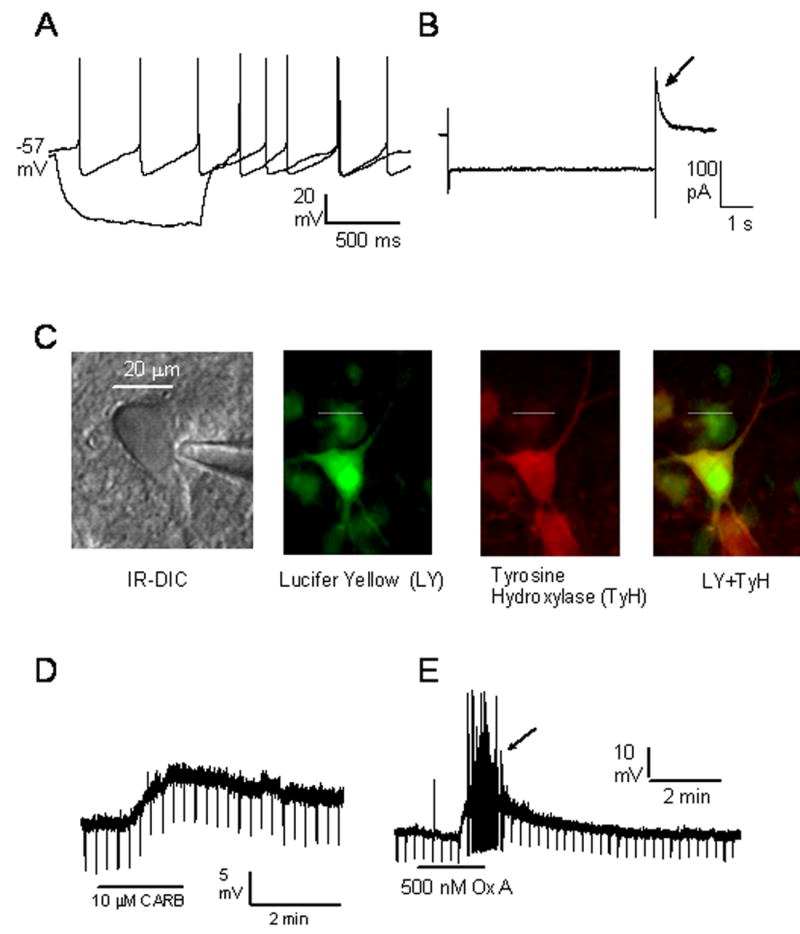
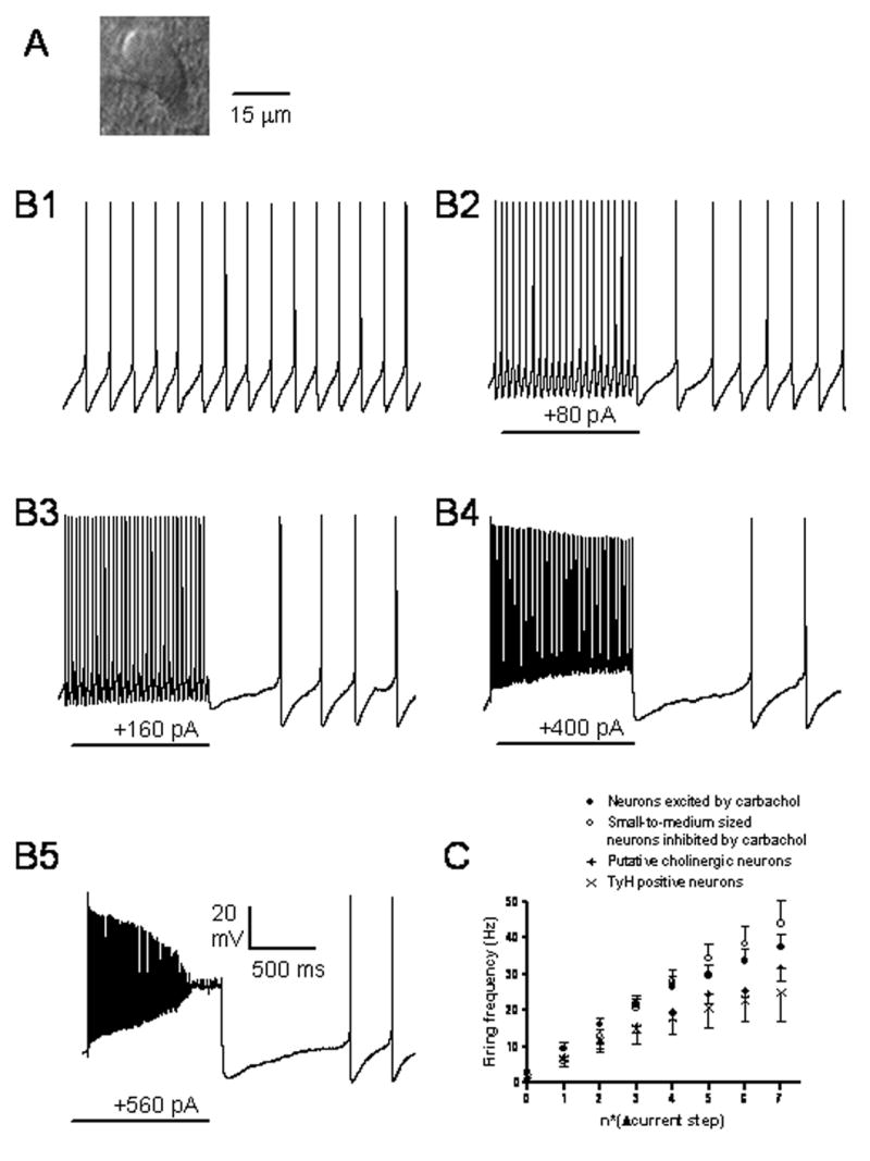
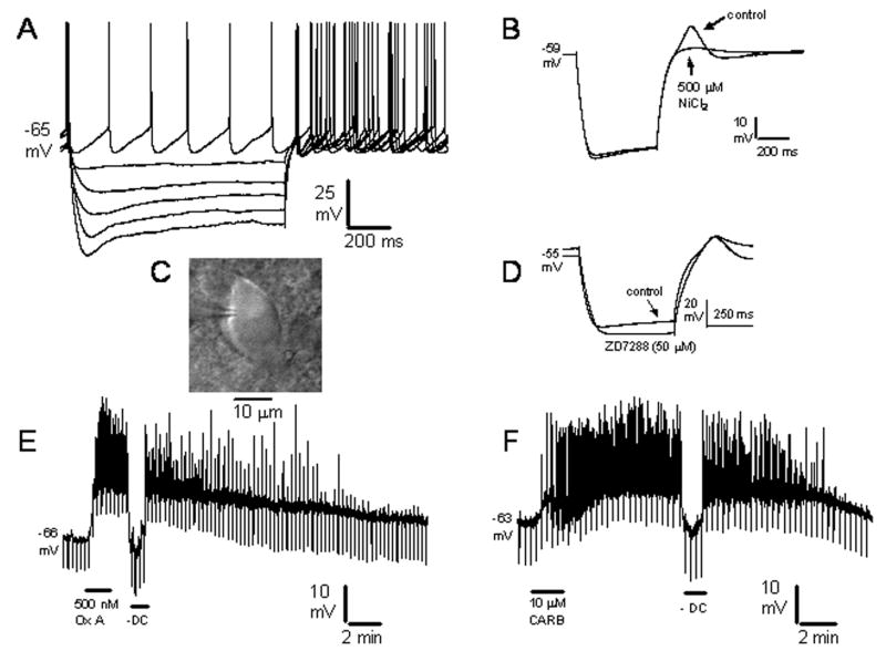
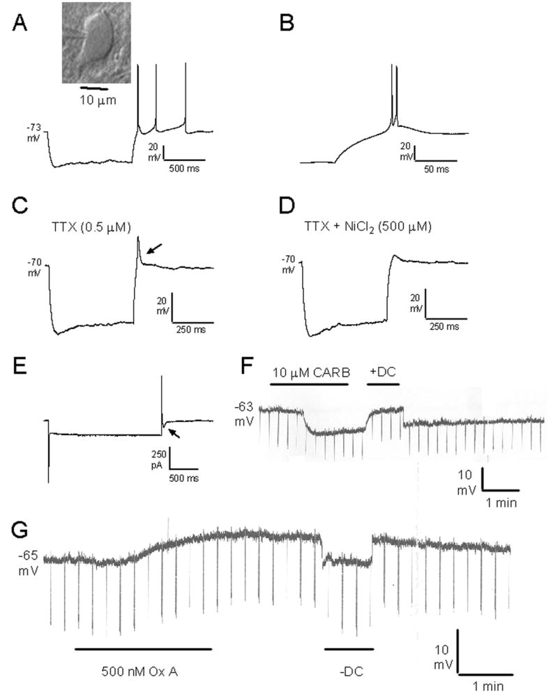
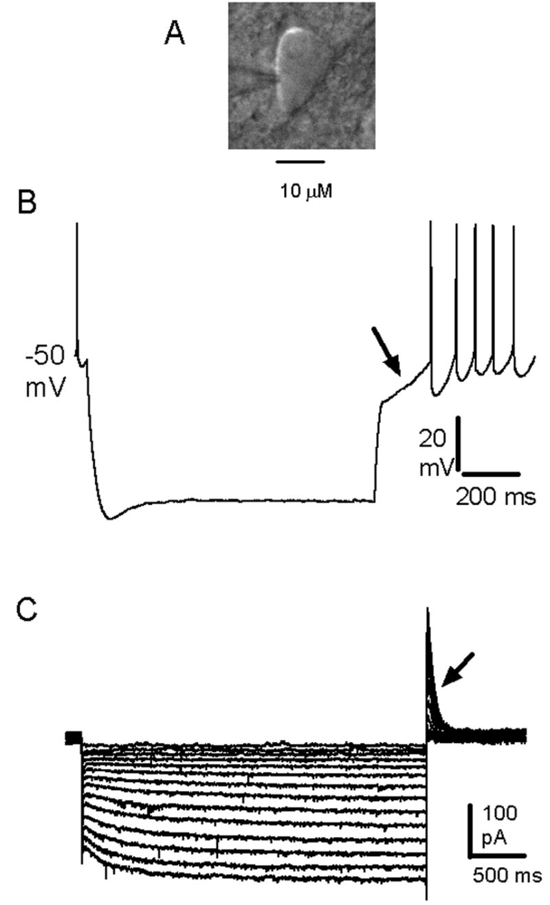
Pharmacological, lesion and single-unit recording techniques in several animal species have identified a region of the pontine reticular formation (subcoeruleus, SubC) just ventral to the locus coeruleus as critically involved in the generation of rapid-eye-movement (REM) sleep. However, the intrinsic membrane properties and responses of SubC neurons to neurotransmitters important in REM sleep control, such as acetylcholine and orexins/hypocretins, have not previously been examined in any animal species and thus were targeted in this study. We obtained whole-cell patch-clamp recordings from visually identified SubC neurons in rat brain slices in vitro. Two groups of large neurons (mean diameter 30 and 27 mum) were tentatively identified as cholinergic (rostral SubC) and noradrenergic (caudal SubC) neurons. SubC reticular neurons (non-cholinergic, non-noradrenergic) showed a medium-sized depolarizing sag during hyperpolarizing current pulses and often had a rebound depolarization (low-threshold spike, LTS). During depolarizing current pulses they exhibited little adaptation and fired maximally at 30-90 Hz. Those SubC reticular neurons excited by carbachol (n=27) fired spontaneously at 6 Hz, often exhibited a moderately sized LTS, and varied widely in size (17-42 mum). Carbachol-inhibited SubC reticular neurons were medium-sized (15-25 mum) and constituted two groups. The larger group (n=22) was silent at rest and possessed a prominent LTS and associated one to four action potentials. The second, smaller group (n=8) had a delayed return to baseline at the offset of hyperpolarizing pulses. Orexins excited both carbachol excited and carbachol inhibited SubC reticular neurons. SubC reticular neurons had intrinsic membrane properties and responses to carbachol similar to those described for other reticular neurons but a larger number of carbachol inhibited neurons were found (>50%), the majority of which demonstrated a prominent LTS and may correspond to pontine-geniculate-occipital burst neurons. Some or all carbachol-excited neurons are presumably REM-on neurons.


Figures

Figure 1
Figure 1
Neuroanatomy of the rat subcoeruleus (SubC) region. Left panels: Schematics of the rostral (top, −9.30 mm with respect to Bregma) and caudal (bottom, −9.68 mm with respect to Bregma) SubC regions (SubCD + SubCA) adapted from Paxinos and Watson (1998). Black dots represent very large, mesencephalic (Me5) neurons located lateral to the LDTg and LC. Top right: Distribution of mesopontine cholinergic neurons at the level of the rostral SubC revealed using staining for neuronal nitric oxide synthase (nNOS) and diaminobenzidine (DAB) as chromogen. Large, darkly stained cholinergic neurons (nNOS positive) are densely concentrated dorsally in the laterodorsal region (LDTg) in the central gray and are also present more sparsely in the SubC region, ventral to the LDTg. Bottom right: Distribution of mesopontine noradrenaline neurons at the level of the caudal SubC revealed using staining for tyrosine hydroxylase (TyH) with diaminobenzidine (DAB) as chromogen. Large, darkly stained noradrenaline neurons (TyH positive) are present dorsally in the LC. Scattered TyH positive neurons are also present in the SubC region, ventral to the LC. Abbreviations: CG, central gray; DMTg, dorsomedial tegmental area; DRC, dorsal raphe, caudal part; DTg, dorsal tegmental nucleus of Gudden; LC, locus coeruleus; LDTg, laterodorsal tegmental nucleus; Me5, mesencephalic 5 nucleus; mlf, medial longitudinal fasciculus; SubCA, subcoeruleus alpha part; SubCD, subcoeruleus, dorsal part.
Figure 2
Figure 2
Properties of large, putative cholinergic neurons in the subcoeruleus region. A: Response to hyperpolarizing and depolarizing current injection in current-clamp. B: Response to hyperpolarizing current injection in the presence of the voltage-gated sodium channel blocker tetrodotoxin. C, D: Continuous recordings of membrane potential in the presence of tetrodotoxin (0.5 μM) showing the hyperpolarization caused by carbachol (CARB, C) and the depolarization caused by orexin A (Ox A, D). Depolarizing (+DC) or hyperpolarizing current injection (-DC) was used to return the membrane potential to the baseline in order to assess the effects of the drugs on input resistance, as assessed by the voltage responses to short (500 ms) hyperpolarizing current steps (downward deflections). E: Slow voltage ramps from −30 to −110 mV reveal that the current induced by the cholinergic agonist carbachol (CARB) is outward at the resting membrane potential (−75 mV) and reverses around the equilibrium potential for potassium (−100 mV). F: Infra-red differential interference contrast (IR-DIC) image of the recording pipette and neuron for one neuron of this class whose responses are shown in A, B and E.
Figure 3
Figure 3
Properties of tyrosine hydroxylase (TyH) positive neurons in the subcoeruleus region. A: Response to hyperpolarizing current injection and spontaneous firing in current-clamp. B: Response to a hyperpolarizing voltage step in voltage-clamp in the presence of the voltage-gated sodium channel blocker tetrodotoxin. Note the transient outward current (arrow) at the offset of the step. C: Infra-red differential interference contrast (IR-DIC) image of the recording pipette and neuron for one neuron of this class whose responses are shown in A, B. This same neuron was filled with the dye Lucifer Yellow. Post-hoc staining revealed that this neuron was TyH positive. D, E: Continuous recordings of membrane potential in the presence of tetrodotoxin (0.5 μM) showing the depolarizations caused by carbachol (CARB, D) and orexin A (Ox A, E). Input resistance was assessed by the voltage responses to short (500 ms) hyperpolarizing current steps (downward deflections).
Figure 4
Figure 4
Response of subcoerulean (SubC) reticular neurons to depolarizing current injection. A: Infra-red differential interference contrast (IR-DIC) image of the neuron with the responses depicted in B. B1: this neuron fired spontaneously in the absence of injected current. B2-B5: response to increasing current injection. SubC neurons increased showed increased firing rate without adaptation. During large current pulses inactivation of voltage-gated sodium channels occurred leading to smaller action potentials and/or cessation of firing (B4, B5). Prolonged afterhyperpolarizations proportional to the size of the current step which delayed resumption of firing are also visible at the offset of the depolarizing current injection. C: Current frequency plots for different types of neurons found in the SubC. The Δcurrent step value was one fifth of the amount of current needed to hyperpolarize the neuron to −120 mV.
Figure 5
Figure 5
Properties of SubC reticular neurons excited by carbachol (CARB-E neurons). A: Responses to a series of hyperpolarizing current pulses and spontaneous firing in current-clamp. B: Response to a hyperpolarizing current step in the presence of the voltage-gated sodium channel blocker tetrodotoxin (TTX). Note the rebound depolarization at the offset of the step which is blocked by the calcium channel antagonist nickel chloride (arrow). C: Infrared differential contrast (IR-DIC) image of the recording pipette and neuron for one neuron of this class whose responses are shown in A, B, D-F. D: Response to hyperpolarizing current in the presence of the voltage-gated sodium channel blocker tetrodotoxin. Note the depolarizing sag during the step which is blocked by the H-current blocker ZD7288. E, F: Continuous recordings of membrane potential in the presence of tetrodotoxin (0.5 μM) showing the depolarizations caused by orexin A (Ox A, E) and carbachol (CARB, F). Note the high-amplitude calcium spikes (upward deflections) at the peaks of the depolarizations caused by orexin A or carbachol. Hyperpolarizing current injection (-DC) was used to return the membrane potential to the baseline in order to assess the effects of the drugs on input resistance, as assessed by the voltage responses to short (500 ms) hyperpolarizing current steps (downward deflections).
Figure 6
Figure 6
Properties of SubC reticular neurons inhibited by carbachol and possessing a large low-threshold spike (CARB-I LTS neurons). A: Response to hyperpolarizing current injection in current-clamp. Inset: Infra-red differential interference contrast (IR-DIC) image of the recording pipette and neuron for one neuron of this class. B: Expanded view of the voltage response at the offset of the hyperpolarizing step shown in A. Note the doublet of action potentials. C: Response to hyperpolarizing current injection in the presence of the voltage-gated sodium channel blocker tetrodotoxin (TTX). Note the large LTS (arrow). D: The LTS is blocked by application of the calcium channel antagonist nickel chloride. E: Current response to a hyperpolarizing voltage step in voltage-clamp. Note the transient inward (T) currrent at the offset of the step (arrow). F, G: Continuous chart recordings of membrane potential in the presence of tetrodotoxin (0.5 μM) showing the hyperpolarization caused by carbachol (CARB, F) and the depolarization caused by orexin A (Ox A, F). Depolarizing (+DC) or hyperpolarizing current injection (-DC) was used to return the membrane potential to the baseline in order to assess the effects of the drugs on input resistance, as assessed by the voltage responses to short (500 ms) hyperpolarizing current steps (downward deflections).
Figure 7
Figure 7
Intrinsic membrane properties of SubC reticular neurons inhibited by carbachol and possessing a transient outward current (CARB-I A-current neurons). A: C: Infra-red differential interference contrast (IR-DIC) image of the recording pipette and neuron for one neuron of this class whose responses are shown in B, C. B: Response to hyperpolarizing current injection. Note the depolarizing sag during the step and the delayed return to baseline at the offset of the step (arrow). C: Responses to a series of hyperpolarizing voltage steps in voltage-clamp in the presence of the voltage-gated sodium channel blocker tetrodotoxin. Note the inward (H) current during the step and the transient outward (A) current (arrow) at the offset of the step (arrow).

Similar articles

Cited by

References

    1. Amzica F, Steriade M. Progressive cortical synchronization of ponto-geniculo-occipital potentials during rapid eye movement sleep. Neuroscience. 1996;72:309–314. - PubMed
    1. Aserinsky E, Kleitman N. Regularly occurring periods of eye motility, and concomitant phenomena, during sleep. Science. 1953;118:273–274. - PubMed
    1. Baghdoyan HA, Rodrigo-Angulo ML, McCarley RW, Hobson JA. Site-specific enhancement and suppression of desynchronized sleep signs following cholinergic stimulation of three brainstem regions. Brain Res. 1984;306:39–52. - PubMed
    1. Baghdoyan HA, Rodrigo-Angulo ML, McCarley RW, Hobson JA. A neuroanatomical gradient in the pontine tegmentum for the cholinoceptive induction of desynchronized sleep signs. Brain Res. 1987;414:245–261. - PubMed
    1. Blanco-Centurion C, Gerashchenko D, Salin-Pascual RJ, Shiromani PJ. Effects of hypocretin2-saporin and antidopamine-beta-hydroxylase-saporin neurotoxic lesions of the dorsolateral pons on sleep and muscle tone. Eur J Neurosci. 2004;19:2741–2752. - PMC - PubMed

Publication types

MeSH terms

LinkOut - more resources