. 2006 Dec;14(6):831-9.
doi: 10.1016/j.ymthe.2006.08.009. Epub 2006 Sep 27.

Autophagy and mistargeting of therapeutic enzyme in skeletal muscle in Pompe disease

Affiliations

Autophagy and mistargeting of therapeutic enzyme in skeletal muscle in Pompe disease

Tokiko Fukuda et al. Mol Ther. 2006 Dec.

Abstract

Enzyme replacement therapy (ERT) became a reality for patients with Pompe disease, a fatal cardiomyopathy and skeletal muscle myopathy caused by a deficiency of glycogen-degrading lysosomal enzyme acid alpha-glucosidase (GAA). The therapy, which relies on receptor-mediated endocytosis of recombinant human GAA (rhGAA), appears to be effective in cardiac muscle, but less so in skeletal muscle. We have previously shown a profound disturbance of the lysosomal degradative pathway (autophagy) in therapy-resistant muscle of GAA knockout mice (KO). Our findings here demonstrate a progressive age-dependent autophagic buildup in addition to enlargement of glycogen-filled lysosomes in multiple muscle groups in the KO. Trafficking and processing of the therapeutic enzyme along the endocytic pathway appear to be affected by the autophagy. Confocal microscopy of live single muscle fibers exposed to fluorescently labeled rhGAA indicates that a significant portion of the endocytosed enzyme in the KO was trapped as a partially processed form in the autophagic areas instead of reaching its target--the lysosomes. A fluid-phase endocytic marker was similarly mistargeted and accumulated in vesicular structures within the autophagic areas. These findings may explain why ERT often falls short of reversing the disease process and point toward new avenues for the development of pharmacological intervention.


Figures

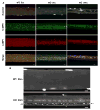
FIG. 1
FIG. 1. Progressive age-dependent autophagic build-up in type II fibers from KO
(A) Representative confocal images of fixed single fibers from EDL muscle. Immunostaining for LC3 (autophagosomal marker) and LAMP1 (late endosomal/lysosomal marker) was performed on myofibers of 1, 5, and 24 month-old KO, and 24 month-old WT. Most of the material in the autophagic area in 24 month-old KO is presumably autofluorescence since it has a broad emission spectrum (see also Fig. 2A and 2B, KO 24m). Note the difference in the fiber size between the WT and KO in old mice. Bars, 10μm. (B) View of the area of autophagic activity in a fiber from EDL muscle of a 1 month-old KO (different fiber than in 1A). Confocal image of a fixed fiber double-stained for LC3 and LAMP1. The merged image shows multiple small LAMP1-positive structures within the LC3-positive autophagosomes. Bar, 2μm.
FIG. 2
FIG. 2. CI-MPR and LAMP 1-positive structures in autophagic areas in KO
(A) Representative confocal images of fixed single fibers from EDL muscle. Immunostaining for CI-MPR (negative marker for lysosomes) and LAMP1 was performed on myofibers from WT (shown for 5 month-old) and KO mice. Nuclei were stained with Hoechst. CI-MPR staining in WT fibers appears as small dot-like structures often overlapping or in close proximity to LAMP1-positive structures. In fibers from 5 month-old KO the receptor is abundant in the autophagic area. In fibers from 24 month-old KO most of the structures within the autophagic areas are autofluorescence with a broad emission spectrum as evidenced by a similar pattern produced by any of the three excitation lasers (405nm, 488nm, and 543nm). (B). Massive accumulation of autofluorescent materials in autophagic area from 24 month-old KO is contrasted to age-matched WT fiber. Note the difference in fiber size. Confocal images of EDL fibers stained with Hoechst were taken using 405nm excitation. The autophagic area, which interrupts muscle striations in the KO fiber, is located toward the periphery of this particular fiber (KO 24m). Bright structures with irregular shape are autofluorescent material in the KO (arrows). Small dark structures (arrowhead) outside the autophagic area are most likely swollen lysosomes. Nuclei, out of focus in this single optical section, appear as a chain in the center of the KO fiber. Bars, 10μm.
FIG. 3
FIG. 3. Vesicular structure of the autophagic areas
(A) Multiple vesicles could be observed in live KO fibers by transmitted light microscopy in the core of the fiber. Live gastrocnemius fibers with satellite cells (arrows) from 2 month-old WT and KO are shown. Bar, 20μm. (B) Confocal image of a live fiber (G of a 3 month-old KO) incubated for 30 min with LysoTracker (75 nM), showing multiple acidic vesicles in the autophagic area. Black arrows point to the margins of the fiber, and white arrows point to the autophagic area in the core of the fiber on the DIC (Differential Interference Contrast) image. Note that Lysotracker is not visible outside the autophagic area because the detection gain was adjusted to avoid saturation in the autophagic area. Bar, 10μm.
FIG. 4
FIG. 4. Detection of the endocytosed fluorescently labeled rhGAA and dextran in autophagic area
(A) Confocal images showing the distribution of the labeled enzyme in a live fiber from EDL of a 3 month-old KO. Labeled rhGAA is concentrated in the autophagic area, which is marked by arrows in the DIC image. The two images on the left were taken by channel mode. The contribution of autofluorescence was evaluated by separation of Alexa Fluor 546-specific emission signal from the autofluorescence with a spectral detector (lambda mode) and linear unmixing analysis. (B) Confocal images showing the distribution of the labeled enzyme in a live fiber from EDL muscle of a 3 month-old WT mouse. The images were taken by channel mode. (C) Confocal images showing the distribution of Alexa Fluor 546-conjugated dextran and contribution of autofluorescence in a live KO fiber from EDL of a 3-month-old mouse. (D) Confocal images showing the distribution of the labeled enzyme and contribution of autofluorescence in a fiber from G muscle of a 3-month-old KO mouse. Unlike the image in (A), the live fiber was first labeled with Alexa Fluor 546-rhGAA and then fixed. The images on the left in (C) and (D) were taken by channel mode. Bars, 10μm.
FIG. 5
FIG. 5. Detection of administered unlabeled rhGAA in whole muscle tissues and fibers
Western blots showing the levels of endogenous GAA in WT and the level of rhGAA in KO after ERT (shown for gastrocnemius muscle). T: whole muscle tissue, F: fibers. In (A) protein-A purified anti-human GAA antibody was used. The levels of the precursor and the processed forms in WT fibers, but not in the KO fibers on ERT, are similar to those in whole tissue lysates. In (B) affinity purified anti-human GAA antibody was used. This antibody recognized the 110 kDa administered rhGAA (0.05ng), thus confirming its specificity. GAPDH was used as a loading control.

References

    1. Sly WS. Enzyme replacement therapy for lysosomal storage disorders: successful transition from concept to clinical practice. Mo Med. 2004;101:100–104. - PubMed
    1. Hirschhorn R, Reuser AJ. Glycogen Storage Disease Type II: Acid alpha-Glucosidase (Acid Maltase) Deficiency. In: Seriver CR, Beaudet AL, Sly WS, Valle D, editors. The metabolic and molecular basis of inherited disease. McGraw-Hill; New York: 2001. pp. 3389–3420.
    1. Van den Hout, et al. The natural course of infantile Pompe's disease: 20 original cases compared with 133 cases from the literature. Pediatrics. 2003;112:332–340. - PubMed
    1. Kishnani PS, Hwu WL, Mandel H, Nicolino M, Yong F, Corzo D. A retrospective, multinational, multicenter study on the natural history of infantile-onset Pompe disease. J Pediatr. 2006;148:671–676. - PubMed
    1. Hagemans ML, et al. Clinical manifestation and natural course of late-onset Pompe's disease in 54 Dutch patients. Brain. 2005;128:671–677. - PubMed

Publication types

MeSH terms