. 2006 Nov;129(Pt 11):2945-56.
doi: 10.1093/brain/awl254. Epub 2006 Sep 28.

Structural anatomy of empathy in neurodegenerative disease

Affiliations

Structural anatomy of empathy in neurodegenerative disease

Katherine P Rankin et al. Brain. 2006 Nov.

Abstract

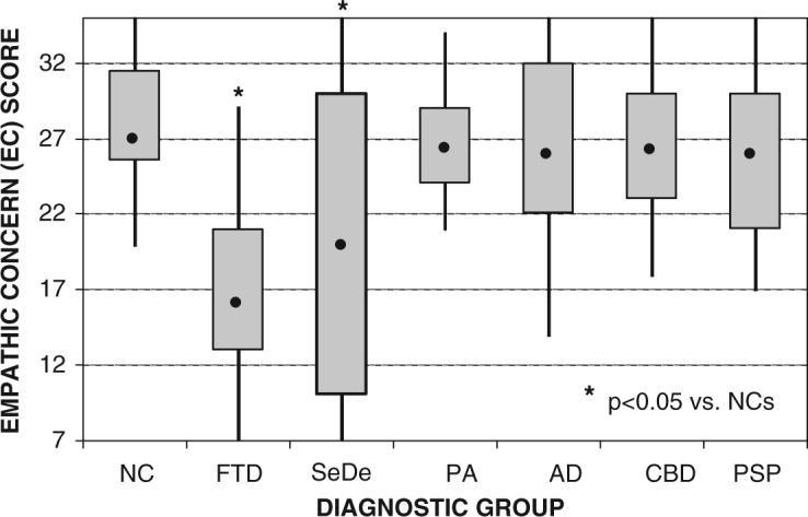
Empathy is a complex social behaviour mediated by a network of brain structures. Recently, several functional imaging studies have investigated the neural basis of empathy, but few corroborative human lesion studies exist. Severe empathy loss is a common feature of frontotemporal lobar degeneration (FTLD), and is also seen in other neurodegenerative diseases. In this study, the neuroanatomic basis of empathy was investigated in 123 patients with FTLD, Alzheimer's disease, corticobasal degeneration and progressive supranuclear palsy using the Interpersonal Reactivity Index (IRI). IRI Empathic Concern and Perspective taking scores were correlated with structural MRI brain volume using voxel-based morphometry. Voxels in the right temporal pole, the right fusiform gyrus, the right caudate and right subcallosal gyrus correlated significantly with total empathy score (P < 0.05 after whole-brain correction for multiple comparisons). Empathy score correlated positively with the volume of right temporal structures in semantic dementia, and with subcallosal gyrus volume in frontotemporal dementia. These findings are consistent with previous research suggesting that a primarily right frontotemporal network of brain regions is involved in emotion processing, and highlights the roles of the right temporal pole and inferior frontal/striatal regions in regulating complex social interactions. This is the first large-scale lesion study to investigate the neural basis of empathy using correlational analytic methods. The results suggest that the right anterior temporal and medial frontal regions are essential for real-life empathic behaviour.


Figures

Fig. 1
Fig. 1
Boxplots of EC score classified by the six diagnostic groups compared with an age-matched healthy comparison group of 25 subjects (NC = normal controls; FTD = frontotemporal dementia variant of FTLD; SeDe = semantic dementia variant of FTLD; PNFA = progressive non-fluent aphasia variant of FTLD; AD = Alzheimer's disease; CBD = corticobasal degeneration; PSP = progressive supranuclear palsy). The dot inside of each plot represents the group's median score. The box represents the range of scores for the group's second and third quartiles. The whiskers represent the score range for the top and bottom quartiles. Group comparisons were performed using a post hoc Dunnett–Hsu test controlling for sex and age.
Fig. 2
Fig. 2
Boxplots of PT score classified by the six diagnostic groups compared with an age-matched healthy comparison group of 20 subjects (NC = normal controls; FTD = frontotemporal dementia variant of FTLD; SeDe = semantic dementia variant of FTLD; PNFA = progressive aphasia variant of FTLD; AD = Alzheimer's disease; CBD = corticobasal degeneration; PSP = progressive supranuclear palsy). The dot inside of each plot represents the group's median score. The box represents the range of scores for the group's second and third quartiles. The whiskers represent the score range for the top and bottom quartiles. Group comparisons were performed using a post hoc Dunnett–Hsu test controlling for sex and age.
Fig. 3
Fig. 3
Continuous, unthresholded map of t-scores for empathy total score analysis across all diagnostic groups, superimposed on a normal control template image (SPM2: single_subj_T1.mnc). The figure represents axial slices taken every 3 mm from z = −51 to z = +18. t-Scores range from 2.00 (blue–black) to 5.91 (red), and include slices from all three clusters containing significant voxels in the analysis.
Fig. 4
Fig. 4
Scatterplot of total empathy score (EC + PT) versus voxel intensity at the most significant voxel in the right anterior temporal lobe cluster (58, 10, −33). Voxel intensities were extracted from the SPM analysis after adjustment for age, sex and total intracranial volume.
Fig. 5
Fig. 5
(A) Main effect of EC score, showing rendered, sagittal (x = 7) and axial (z = 0) views of voxels significantly related to EC score at P < 0.001 uncorrected for multiple comparisons across the whole brain. Maps of significant correlation were superimposed on sections of a normal brain template image (SPM2: single_subj_T1.mnc). The design matrix for this analysis contained only EC score, with sex, age and TIV included as nuisance covariates, and a t-test was used. (B) Main effect of PT score, showing voxels significantly related to PT score at P < 0.001 uncorrected. The design matrix for this analysis contained only PT score, with sex, age and TIV included as nuisance covariates, and a t-test was used.

References

    1. Adolphs R. Social cognition and the human brain. Trends Cogn Sci. 1999;3:469–79. - PubMed
    1. Adolphs R, Damasio H, Tranel D, Cooper G, Damasio AR. A role for somatosensory cortices in the visual recognition of emotion as revealed by three-dimensional lesion mapping. J Neurosci. 2000;20:2683–90. - PMC - PubMed
    1. Allison T, Puce A, McCarthy G. Social perception from visual cues: role of the sts region. Trends Cogn Sci. 2000;4:267–78. - PubMed
    1. Anderson SW, Bechara A, Damasio H, Tranel D, Damasio AR. Impairment of social and moral behavior related to early damage in human prefrontal cortex. Nat Neurosci. 1999;2:1032–7. - PubMed
    1. Beer JS, Heerey E, Keltner D, Scabini D, Knight R. The regulatory function of self-conscious emotion: insights from patients with orbitofrontal damage. J Pers Soc Psychol. 2003;85:594–604. - PubMed

Publication types

MeSH terms