. 2006 Dec 1;577(Pt 2):641-58.
doi: 10.1113/jphysiol.2006.118711. Epub 2006 Sep 28.

Modelling spinal circuitry involved in locomotor pattern generation: insights from the effects of afferent stimulation

Affiliations

Modelling spinal circuitry involved in locomotor pattern generation: insights from the effects of afferent stimulation

Ilya A Rybak et al. J Physiol. .

Abstract

A computational model of the mammalian spinal cord circuitry incorporating a two-level central pattern generator (CPG) with separate half-centre rhythm generator (RG) and pattern formation (PF) networks has been developed from observations obtained during fictive locomotion in decerebrate cats. Sensory afferents have been incorporated in the model to study the effects of afferent stimulation on locomotor phase switching and step cycle period and on the firing patterns of flexor and extensor motoneurones. Here we show that this CPG structure can be integrated with reflex circuits to reproduce the reorganization of group I reflex pathways occurring during locomotion. During the extensor phase of fictive locomotion, activation of extensor muscle group I afferents increases extensor motoneurone activity and prolongs the extensor phase. This extensor phase prolongation may occur with or without a resetting of the locomotor cycle, which (according to the model) depends on the degree to which sensory input affects the RG and PF circuits, respectively. The same stimulation delivered during flexion produces a temporary resetting to extension without changing the timing of following locomotor cycles. The model reproduces this behaviour by suggesting that this sensory input influences the PF network without affecting the RG. The model also suggests that the different effects of flexor muscle nerve afferent stimulation observed experimentally (phase prolongation versus resetting) result from opposing influences of flexor group I and II afferents on the PF and RG circuits controlling the activity of flexor and extensor motoneurones. The results of modelling provide insights into proprioceptive control of locomotion.


Figures
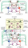
Figure 1
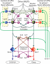
Figure 1. Reflex circuitry of group I afferents and its reorganization during locomotion
A, the simplified schematic diagram of the neural circuitry of spinal reflexes operating under non-locomotor conditions. B, reorganization of the spinal circuitry during (fictive) locomotion. Populations of interneurones are represented by spheres. Excitatory and inhibitory synaptic connections are shown by arrows and small circles, respectively. Populations of flexor (Mn-F) and extensor (Mn-E) motoneurones are represented by diamonds. Excitatory tonic locomotor drives are shown in grey. Connections of group I (Ia and Ib) sensory afferents are in green. In B, the upper area enclosed by dashed lines indicates the CPG circuits including the rhythm generator (RG) and pattern formation (PF) network. The full description of CPG architecture and performance can be found in the accompanying paper, Rybak et al. (2006). During the extensor phase of fictive locomotion, the Iab-E population (blue) is released from inhibition by the In-E population (grey) and mediates phase-dependent disynaptic excitation of extensor motoneurones by group I extensor afferents. During both locomotor phases, the In population (grey) inhibits both the Ib-E and Ib-F populations and hence suppresses non-reciprocal inhibition. See text for details and definitions.
Figure 2
Figure 2
Model schematic diagram of the spinal cord circuitry integrated with the locomotor CPG used for simulation of the effects of extensor group I and cutaneous afferent stimulation during fictive locomotion The non-reciprocal inhibition illustrated in Fig. 1 has been removed and interneurone populations Irg-E and Ipf-E (both blue) have been added to mediate the access of sensory information from extensor group I (Ia and Ib, green) and cutaneous (Cut, brown) afferents to the rhythm generator (RG-E) and the pattern formation (PF-E) networks. See text for details and definitions. The Iab-E population mediates phase-dependent disynaptic excitation of extensor motroneurons.
Figure 3
Figure 3. Modelling the effects of group I extensor afferent stimulation delivered during flexion
A, the stimulation applied to group Ia and Ib afferents is shown in the top trace. Other traces show activities of the neural populations in Fig. 2 represented by the average histograms of firing frequency (number of spikes per second per neurone, bin = 30 ms). Stimulus amplitude (dmax) was 0.8. The tonic drives to RG-F (drg-f) and RG-E (drg-e) populations were 0.57 and 0.39, respectively. Note that here and in the subsequent figure legends, each subscripted variable d defines the normalized value representing the corresponding particular drive or afferent input, for details see eqn (10) in the preceding paper by Rybak et al. (2006). B, an example of the effect of stimulating extensor group I afferents in the lateral gastrocnemius and soleus (LGS) nerve (1.6T, where T is the threshold for activation of group I afferents; 20 shocks, 100 Hz, filled rectangle) during the flexion phase of MLR-evoked fictive locomotion (modified from Fig. 6A in Guertin et al. 1995). Stimulation produced a premature onset of extension as seen in the activities of the rectified and integrated hip (anterior biceps, AB) and knee (quadriceps, Quad) extensor electroneurogram (ENG) recordings and terminated the ongoing flexion phase (tibialis anterior, TA, an ankle flexor). Note in both A and B, the stimulation resulted in a short extensor burst that was followed by a shortened flexion phase so that the timing of the following step cycles did not change (i.e. the locomotor rhythm was not reset; see arrows at the bottom showing equal locomotor periods before, during and after application of stimulation).
Figure 4
Figure 4. Modelling the effects of group I extensor afferent stimulation during extension
Aa and Ab, examples of modelling the effects of stimulation of the group I extensor afferents during extension (see text for details). The applied stimuli are shown in the top traces. The stimulus amplitude (dmax) was 0.8 in Aa and 3.2 in Ab. In Aa, drives to RG-F and RG-E populations were: 0.52 and 0.43, respectively; and in Ab were: 0.52 and 0.47, respectively. Ba and Bb, the effects of stimulation of extensor group I afferents during MLR-evoked fictive locomotion. In Ba, stimulation of plantaris (Pl) group I afferents during extension (1.6 times threshold (1.6T), 20 shocks, 100 Hz; adapted from Fig. 1A in Guertin et al. 1995) increased the size and duration of extensor motoneurone activity (medial gastrocnemius, MG) and shortened the duration of the following flexor phase as seen in the sartorius (Sart) ENG. Note that in both Aa and Ba, the duration of each flexion phase following the prolonged extension phase was shortened so that the locomotor periods did not change. The locomotor rhythm was not reset (see equal length arrows at the bottom). In panel Bb, hip extensor (semimembranosus, Sm) muscle afferents were electrically stimulated during extension (2T, 20 shocks, 100 Hz). This figure is from unpublished observations obtained during the experiments reported by Guertin et al. (1995). In contrast to the results shown in Aa and Ba, in Ba and Bb the flexion phase that follows the stimulus-evoked extension phase prolongation (see MG trace in Bb) was not shortened (see TA trace) and the step cycle period increased with each stimulus delivery (see arrows at the bottom).
Figure 5
Figure 5. Modelling the effects of cutaneous nerve stimulation
Aa and Ab, using the schematic diagram shown in Fig. 2, sensory stimuli were applied to the cutaneous afferents during the flexor (Aa) and extensor (Ab) phases of locomotion. The applied stimulation affected both the PF and RG levels of CPG (on the extensor side, see Fig. 2). The applied stimuli are shown in the top traces (dmax= 3.0). Drives to RG-F and RG-E populations were: 0.43 and 0.53, respectively. Ba and Bb, experimentally recorded effects of stimulation of a cutaneous nerve during flexion and extension. The stimulations (20 shocks, 100 Hz) were applied to the distal portion of the tibial nerve. Stimulation applied during flexion (Ba) terminated the ongoing flexor phase (sartorius, Sart) and produced a premature onset of extension (seen in activities of anterior biceps (AB) and lateral gastrocnemius combined with soleus (LGS)). Stimulation applied during extension (Bb) prolonged the extension phase (seen in activities of semimembranosus (Sm) and medial gastrocnemius (MG)). The recordings are from Figs 7C and D of Guertin et al. (1995). Note that in Aa and Ba, the afferent stimulation shortened the cycle period, whereas in Ab and Bb, cycle period was prolonged (see arrows at the bottom of each part of the figure).
Figure 6
Figure 6. Schematic diagram of the model used for simulation of flexor afferent stimulation
Flexor group I afferents access the RG and PF elements on the flexor side of the CPG through interneurone populations (Irg-F and Ipf-F, respectively, both are shown red). Flexor group II afferents project to the extensor side of the CPG via the Irg-E and Ipf-E populations, respectively (shown blue). See text for details and definitions.
Figure 7
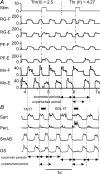
Figure 7. Modelling the effects of stimulation of flexor afferents during flexion
A, the applied stimuli are shown in the top trace (stimulus amplitude (dmax= 5.0). The first stimulus terminated ongoing flexion and initiated a premature switch to extension. At the 5.5 s time point in the simulation (i.e. before the second stimulus application), the threshold of group II flexor afferent activation (Thr(II), see eqn (1)) was increased from 2.5 to 4.27 (see labels at the top). As a result, group II afferent action on the extensor CPG circuitry was reduced and the second stimulus produced a prolongation of the ongoing flexion phase (see PF-F and Mn-F activity). Note that both stimulus presentations reset the locomotor rhythm (see RG-E and RG-F traces). Tonic drives to RG-F (drg-f) and RG- E (drg-e) populations were: 0.55 and 0.53, respectively. B, experimental effects of five times threshold (5T) stimulation of flexor afferents during flexion. Stimulation of tibialis anterior (TA) afferents terminated the flexion phase (sartorius, Sart and peroneus longus, PerL) and advanced the onset of extensor (semimembranosus combined with anterior biceps, SmAB and combined lateral and medial gastrocnemius and soleus nerves, GS) activity. Stimulation of another flexor nerve (extensor digitorum longus, EDL) in the same run of fictive locomotion prolonged the ongoing flexion phase (see activities of Sart and PerL). Data from Stecina et al. (2005). In both the simulated and experimental trials, the locomotor rhythm was affected by flexor nerve stimulation with the first stimulation shortening, and the second prolonging, the ongoing locomotor period (see arrows at the bottom of A and B).
Figure 8
Figure 8. Modelling the variable effects evoked by flexor muscle afferent stimulation during flexion on locomotor output
AaAe, results from modelling experiments illustrating variable effects produced by the activation of flexor afferents (filled rectangles) during the flexion phase of locomotion. All model parameters and parameters of stimulation were the same as in Fig. 7A, except for the threshold of group II flexor afferent activation Thr(II) (see eqn (1)), which was sequentially decreased from Aa to Ae (see values of the Thr(II) at the top of each panel). The filled rectangles at the top indicate the timing and duration of the stimuli. Note that the afferent stimulation resets the RG in all simulations except for Aa (see the top and second traces showing RG-F and RG-E population activities). In Ab and Ac, the concomitant activation of PF-F by flexor group I afferents (see Fig. 6) prevented resetting at the PF level (third and fourth traces in Ab and Ac) and the level of motoneurones (bottom two traces). The result therefore was a prolongation of the flexion phase. In Ad, the applied stimulus produced only a short break in the middle of flexor activity and a small, short extensor burst (Mn-E). In Ae, with the lowest Thr(II) (i.e. strongest group II actions), sensory activation produced a full resetting to extension (see text for details). BaBe, experimentally recorded effects of five times threshold (5T) stimulation of extensor digitorum longus (EDL) afferents during the flexion phase of MLR-evoked fictive locomotion. Averaged ENG records during cycles in which stimulation was applied (perturbed cycle) are shown in bold with control (unperturbed) cycles shown directly below. These observations were selected from different experiments (McCrea, 2001; Stecina et al. 2005) and formed the basis of the simulations shown in AaAe. Note the variety of effects observed following 5T EDL stimulation in the different experiments. In Ba, EDL stimulation resulted in a slight increase in flexor (sartorius, Sart) activity with no effect on the timing of flexor or extensor motoneurone bursts. In Bb, there was a prolongation of the ongoing flexor phase and a delay in the activation of extensors. In Bc and Bd, flexor afferent stimulation also prolonged the flexion phase (see Sart in Bc and psoas (Psoas) in Bd) but there was also a brief reduction in flexor motoneurone activity in the middle of the flexor phase and a corresponding small but visible activation of extensors (see the hint of extensor activity in GS in Bc). Both these effects presumably indicate a short lived resetting of flexor activity. These experimental observations are similar to the modelled trials shown in Ac and Ad. Be, shows an example of a rarely observed effect of EDL stimulation in which flexion was terminated and a premature onset of extension initiated. This was another stimulus trial from the same run of fictive locomotion as shown in Bd and is an example of a spontaneous reversal of EDL actions (Stecina et al. 2005).

Similar articles

Cited by

References

    1. Angel MJ, Guertin P, Jiminez I, McCrea DA. Group I extensor afferents evoke disynaptic EPSPs in cat hindlimb extensor motoneurones during fictive locomotion. J Physiol. 1996;494:851–861. - PMC - PubMed
    1. Angel MJ, Jankowska E, McCrea DA. Candidate interneurones mediating group I disynaptic EPSPs in extensor motoneurones during fictive locomotion in the cat. J Physiol. 2005;563:597–610. - PMC - PubMed
    1. Booth V, Rinzel J, Kiehn O. Compartmental model of vertebrate motoneurons for Ca2+-dependent spiking and plateau potentials under pharmacological treatment. J Neurophysiol. 1997;78:3371–3385. - PubMed
    1. Brink E, Jankowska E, McCrea DA, Skoog B. Inhibitory interactions between interneurones in reflex pathways from group Ia and group Ib afferents in the cat. J Physiol. 1983;343:361–373. - PMC - PubMed
    1. Buford JA, Smith JL. Adaptive control for backward quadrupedal walking. III. Stumbling corrective reactions and cutaneous reflex sensitivity. J Neurophysiol. 1993;70:1102–1114. - PubMed

Publication types