. 2007 Feb 1;109(3):1147-55.
doi: 10.1182/blood-2006-02-001339. Epub 2006 Sep 28.

PI3K regulates pleckstrin-2 in T-cell cytoskeletal reorganization

Affiliations

PI3K regulates pleckstrin-2 in T-cell cytoskeletal reorganization

Tami L Bach et al. Blood. .

Abstract

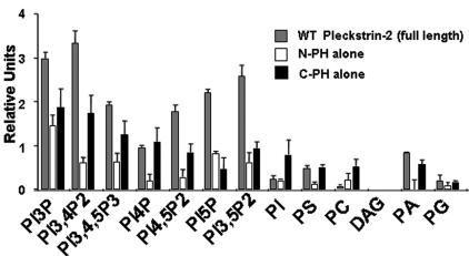
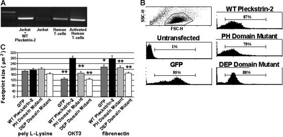
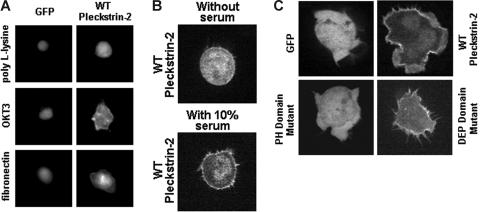
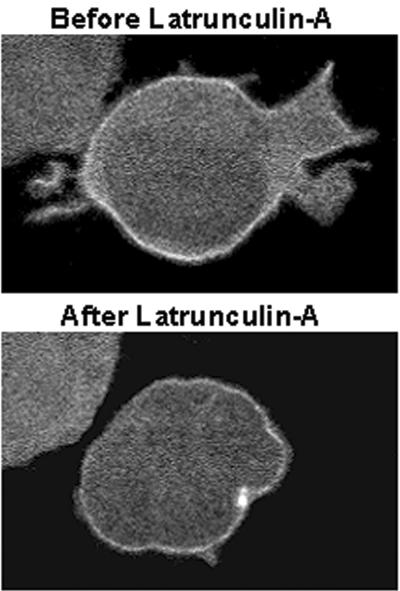
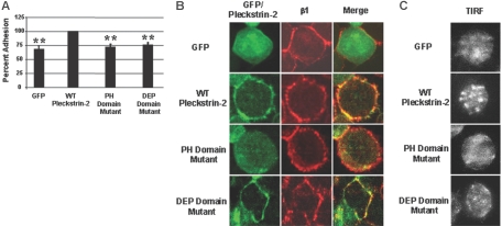
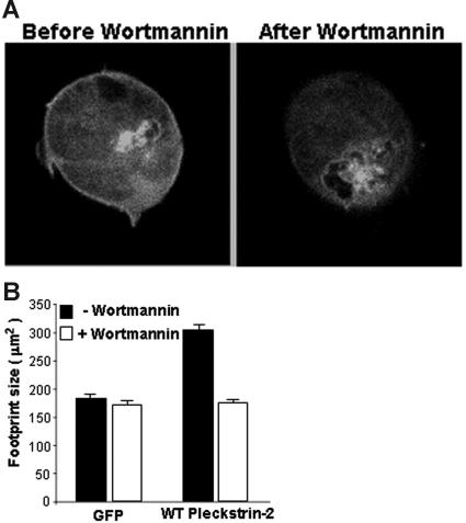
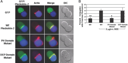
Pleckstrin-2 is composed of 2 pleckstrin homology (PH) domains and a disheveled-Egl-10-pleckstrin (DEP) domain. A lipid-binding assay revealed that pleckstrin-2 binds with greatest affinity to D3 and D5 phosphoinositides. Pleckstrin-2 expressed in Jurkat T cells bound to the cellular membrane and enhanced actin-dependent spreading only after stimulation of the T-cell antigen receptor or the integrin alpha4beta1. A pleckstrin-2 variant containing point mutations in both PH domains failed to associate with the Jurkat membrane and had no effect on spreading under the same conditions. Although still membrane bound, a pleckstrin-2 variant containing point mutations in the DEP domain demonstrated a decreased ability to induce membrane ruffles and spread. Pleckstrin-2 also colocalized with actin at the immune synapse and integrin clusters via its PH domains. Although pleckstrin-2 can bind to purified D3 and D5 phosphoinositides, the intracellular membrane association of pleckstrin-2 and cell spreading are dependent on D3 phosphoinositides, because these effects were disrupted by pharmacologic inhibition of phosphatidylinositol 3-kinase (PI3K). Our results indicate that pleckstrin-2 uses its modular domains to bind to membrane-associated phosphatidylinositols generated by PI3K, whereby it coordinates with the actin cytoskeleton in lymphocyte spreading and immune synapse formation.


Figures

Figure 1
Figure 1
Pleckstrin-2 shows affinity for phosphoinositides with D3 phosphates. GST fusion proteins were made in the pEGX-2TK vector, which allowed the direct (γ-32P) ATP labeling by PKA of the fusion proteins in vitro. Results were viewed by standard autoradiograph techniques and quantified via the Image Quant Program. Relative affinity was calculated by quantitating bound 32P divided by specific activity of the radioactive probe. Shown is the mean ± SEM from 3 independent experiments.
Figure 2
Figure 2
Expression of pleckstrin-2 in Jurkat cells increases cell spreading upon stimulation of cell-surface receptors. (A) RT-PCR products using primers to pleckstrin-2 were derived from Jurkat cells and human T cells and analyzed by agarose electrophoresis. Lane 1, 1 kb ladder; lane 2, Jurkat cells transfected with wild-type (WT) pleckstrin-2; lane 3, untransfected Jurkat cells; lane 4, resting primary human T cells; lane 5, human T cells activated with antibodies to CD3 and CD28. (B) Flow cytometry was performed on Jurkat cells transfected with plasmids that direct the expression of GFP (GFP), GFP fused to the amino-terminus of pleckstrin-2 (WT pleckstrin-2), or GFP fused to the amino-terminus of pleckstrin-2 containing point mutations in both PH domains, N13 N14 N256 (PH domain mutant), or in the DEP domain, N156 N157 N166 (DEP domain mutant). (C) Jurkat cells expressing GFP alone, WT pleckstrin-2, the PH domain mutant, or the DEP domain mutant were plated on either poly-L-Lysine, OKT3, or fibronectin. Images were captured and cell-footprint size was quantified using IP Lab imaging software. Shown is the mean ± SEM of 3 independent experiments. The paired Student t test was performed of the transfectants compared with WT pleckstrin-2 on each matrix. *P < .05; **P < .005.
Figure 3
Figure 3
Expression of pleckstrin-2 in Jurkat cells localizes to the cellular membrane upon stimulation of cell-surface receptors and enhances lamellipodia formation. (A) Jurkat cells expressing GFP alone or GFP-labeled pleckstrin-2 variants were plated upon OKT3 or fibronectin. This figure shows that WT pleckstrin-2 associates with the cell membrane when cells are plated upon OKT3 or fibronectin. Images were captured at ×40 magnification using IP Lab imaging software. (B) Jurkat cells expressing GFP fused to the amino-terminus of wild-type pleckstrin-2 were plated on fibronectin. Real-time confocal imaging of live cells was performed. Shown is a cell expressing pleckstrin-2 before and after addition of 10% FBS. This figure shows that wild-type pleckstrin-2 associates with the membrane and membrane projections rapidly (about 30 seconds) with the addition of serum. Images were captured using an UtraVIEW-LCI confocal scanner at ×100 magnification. (C) Jurkat cells expressing GFP alone or GFP-labeled pleckstrin-2 variants were plated on OKT3. This figure demonstrates that membrane localization is dependent on the pleckstrin-2 PH domains. Similar to WT pleckstrin-2, the DEP domain mutant associates with the cell membrane upon stimulation by the T-cell receptor–activating antibody but does not form membrane ruffles. Images were captured using an UtraVIEW-LCI confocal scanner at × 100 magnification.
Figure 4
Figure 4
Cellular spreading of pleckstrin-2, but not membrane localization, is dependent on the actin cytoskeleton. Jurkat cells expressing GFP fused to the amino-terminus of wild-type pleckstrin-2 were plated on OKT3. When indicated, 1 μM final concentration latrunculin A was added to the chamber slide. Shown is a cell expressing pleckstrin-2 before and after inhibition of the actin cytoskeleton with latrunculin A for 60 seconds. This figure shows that wild-type pleckstrin-2 associates with membrane projections in cells spread on fibronectin, and this membrane localization remains despite the disruption in cellular spreading by the addition of latrunculin A. Similar results were seen when the actin cytoskeleton was disrupted with cytochalasin D. Images were captured using an UtraVIEW-LCI confocal scanner at × 100 magnification.
Figure 5
Figure 5
Expression of pleckstrin-2 in Jurkat cells stimulates clustering of α4β1 and enhances adhesion to fibronectin. (A) Jurkat cells expressing GFP alone, WT pleckstrin-2, the PH domain mutant, or the DEP domain mutant were plated on fibronectin. Jurkat-cell adhesion was quantitated by measuring the activity of acid phosphatase 32. Shown is the mean ± SEM of 6 independent experiments. The paired Student t test was performed of the transfectants compared with WT pleckstrin-2. *P < .005 (B) GFP-fused pleckstrin-2 constructs (green) were plated on fibronectin, fixed, stained with a monoclonal antibody to the integrin β1 subunit (red), and analyzed by indirect immunofluorescence. The merger of the red and green fluorescence to yellow demonstrates that pleckstrin-2 colocalizes with β1. Simultaneous mutations in both PH domains prevented pleckstrin-2 from colocalizing with β1. Images were captured using an UtraVIEW-LCI confocal scanner at ×100 magnification. Data shown are representative of 3 independent experiments. (C) The same GFP-fused pleckstrin-2 constructs, stained with a monoclonal antibody to the integrin β1 subunit, were plated on fibronectin and analyzed using total internal reflection fluorescence (TIRF). Cells transfected with WT pleckstrin-2 demonstrate enhanced β1 clustering at the point of adhesion to fibronectin. Images were captured using a Hamamatsu digital camera and Metamorph software at × 60 magnification. Data shown are representative of 3 independent experiments.
Figure 6
Figure 6
Pleckstrin-2 membrane association and cell spreading are PI3K dependent. (A) Jurkat cells expressing GFP fused to the amino-terminus of wild-type pleckstrin-2 were plated on fibronectin. Real-time confocal imaging of live cells was performed. Shown is a cell expressing pleckstrin-2 before and after inhibition of PI3K with 100 nM wortmannin. This figure shows that wild-type pleckstrin-2 associates with membrane projections in cells spread on fibronectin, and this membrane association is rapidly (about 2 minutes) disrupted by the addition of wortmannin. (B) Jurkat cells expressing GFP (GFP) alone or GFP fused to the amino-terminus of wild-type pleckstrin-2 (WT pleckstrin-2) were plated on fibronectin. Where indicated, 100 nM wortmannin was added for 5 minutes to inhibit PI3K. Images were captured at 100× magnification and cell-footprint size was quantified using IP Lab imaging software. Shown is the mean ± SEM of 3 independent experiments.
Figure 7
Figure 7
Pleckstrin-2 colocalizes with F-actin at the immune synapse. (A) Conjugates were formed between SEE-pulsed Raji B cells stained with CMAC-7 (blue) and Jurkat T cells transfected with the various GFP-fused pleckstrin-2 constructs (green). The fixed conjugates were then stained with rhodamine-phalloidin (red) to label F-actin and analyzed by indirect immunofluorescence at 63× magnification. The merger of the red and green fluorescence to yellow demonstrates that pleckstrin-2 colocalizes with actin at the immune synapse. Simultaneous mutations in both PH domains prevented pleckstrin-2 from colocalizing with actin. Note that the cells are plated on poly-L-lysine and do not demonstrate membrane localization of pleckstrin-2. Data shown are from 1 representative experiment of 3 independent experiments. (B) Colocalization of pleckstrin-2 and actin was quantified by randomly selecting conjugates containing a green T cell contacting a blue B cell. Shown is the mean ± standard deviation of 3 independent experiments. The paired Student t test was performed of WT without SEE stimulation or the pleckstin-2 mutants compared with WT pleckstrin-2. *P < .05; **P < .005.

References

    1. Haslam RJ, Lynham JA, Fox JE. Effects of collagen, ionophore A23187 and prostaglandin E1 on the phosphorylation of specific proteins in blood platelets. Biochem J. 1979;178:397–406. - PMC - PubMed
    1. Tyers M, Rachubinski RA, Stewart MI, et al. Molecular cloning and expression of the major protein kinase C substrate of platelets. Nature. 1988;333:470–473. - PubMed
    1. Ponting CP, Bork P. Pleckstrin's repeat performance: a novel domain in G-protein signaling? Trends Biochem Sci. 1996;21:245–246. - PubMed
    1. Lemmon MA, Ferguson KM, Schlessinger J. PH domains: diverse sequences with a common fold recruit signaling molecules to the cell surface. Cell. 1996;85:621–624. - PubMed
    1. Abrams CS, Zhao W, Belmonte E, Brass LF. Protein kinase C regulates pleckstrin by phosphorylation of sites adjacent to the N-terminal pleckstrin homology domain. J Biol Chem. 1995;270:23317–23321. - PubMed

Publication types