

Review
doi: 10.1136/bmj.38968.683958.AE.
Meningococcal disease and its management in children
Affiliations
- PMID: 17008668
- PMCID: PMC1584345
- DOI: 10.1136/bmj.38968.683958.AE
Item in Clipboard
Review
Meningococcal disease and its management in children
BMJ.
.
No abstract available
Figures
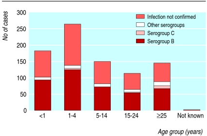
Serogroups of Neisseria meningitidis identified in cases in England and Wales by age: January to March 2005
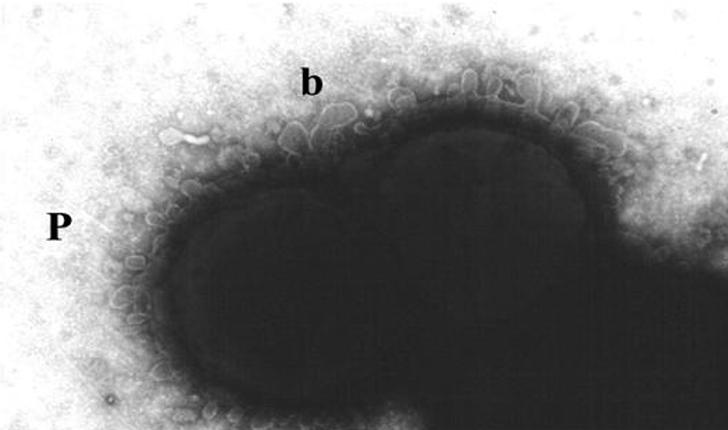
Electronmicrograph showing blebbing (b) of the outer membrane of Neisseria meningitides
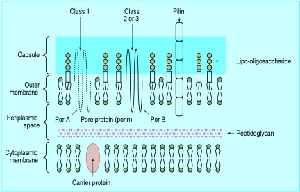
Structure of cell wall of Neisseria meningitidis: 50% of the lipid in the outer leaflet of the outer membrane is lipo-oligosaccharide or endotoxin
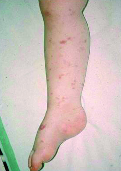
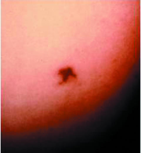
Evolution of non-blanching rash in meningococcal disease. Petechial, purpuric, and vasculitic elements are appearing. The pictures were taken 30 minutes apart. (Reproduced with permission from: Riordan FAI. Factors associated with mortality from meningococcal disease in Merseyside children (MD thesis). Liverpool University, 1995.)
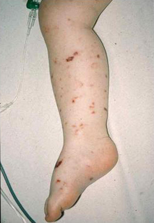
Evolution of non-blanching rash in meningococcal disease. Petechial, purpuric, and vasculitic elements are appearing. The pictures were taken 30 minutes apart. (Reproduced with permission from: Riordan FAI. Factors associated with mortality from meningococcal disease in Merseyside children (MD thesis). Liverpool University, 1995.)
The “tumbler test” is a way of confirming the non-blanching nature of the rash
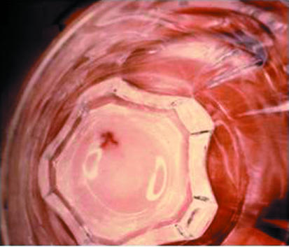
The “tumbler test” is a way of confirming the non-blanching nature of the rash
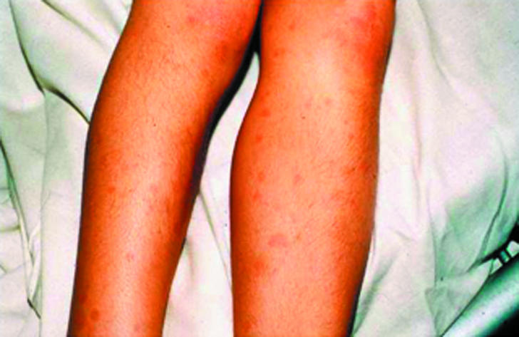
The maculopapular rash of meningococcal disease, indistinguishable from maculopapular rashes in viral infections. (Reproduced with permission from: Marzouk O. Clinical and laboratory aspects of childhood meningococcal disease (MD thesis). Liverpool, 1993.)
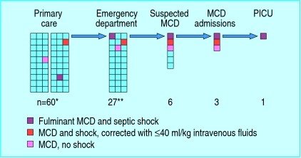
Diagnostic and management pathway from primary care to paediatric intensive care unit (PICU): petechiae in children and meningococcal disease (MCD). (Each square represents one child.) *Range 30-180, but incidence of petechiae in primary care consultations for illness unknown. **Range 27-147, but incidence of petechiae in emergency department varies (Medline search May 2006)
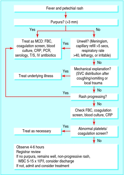
Diagnosis of petechiae in children and meningococcal disease. CRP=C reactive protein; FBC=full blood count; IV=intravenous; MCD=meningococcal disease; PCR=meningococcal polymerase chain reaction; SVC=superior vena cava; T/S=throat swab for meningococci; WBC=white blood cells
Similar articles
-
Meningococcal sepsis.Aust Fam Physician. 2010 May;39(5):276-8. Aust Fam Physician. 2010. PMID: 20485712 Review.
-
Meningococcal septic arthritis of the knee.J R Coll Surg Edinb. 1990 Dec;35(6):398. J R Coll Surg Edinb. 1990. PMID: 2086806 No abstract available.
-
Meningococcal disease--prevention and intervention.Ohio Nurses Rev. 2002 Jul;77(6):15-8; quiz 18-9. Ohio Nurses Rev. 2002. PMID: 15133878 No abstract available.
-
Treating meningococcal infections in children.Hosp Med. 2002 May;63(5):268-73. doi: 10.12968/hosp.2002.63.5.2018. Hosp Med. 2002. PMID: 12066344 Review.
-
New guidelines for management and prevention of meningococcal disease in Australia. Meningococcal Disease Working Party of the National Health and Medical Research Council.Med J Aust. 1997 Jun 2;166(11):598-601. Med J Aust. 1997. PMID: 9201182
Cited by
-
Meningococcemia in an 11 Months Old Infant.Case Rep Infect Dis. 2023 Mar 8;2023:8951318. doi: 10.1155/2023/8951318. eCollection 2023. Case Rep Infect Dis. 2023. PMID: 36936066 Free PMC article.
-
An Adolescent Boy Presenting with Complicated Meningococcal Meningitis Serogroup A: What Is the State of Community Awareness for This Serious Disease?Iran J Public Health. 2014 Sep;43(9):1301-2. Iran J Public Health. 2014. PMID: 26175987 Free PMC article. No abstract available.
-
Nonspecific symptoms dominate at first contact to emergency healthcare services among cases with invasive meningococcal disease.BMC Fam Pract. 2021 Nov 30;22(1):240. doi: 10.1186/s12875-021-01585-8. BMC Fam Pract. 2021. PMID: 34847878 Free PMC article.
-
Multidisciplinary analysis of invasive meningococcal disease as a framework for continuous quality and safety improvement in regional Australia.BMJ Open Qual. 2018 Feb 7;7(1):e000077. doi: 10.1136/bmjoq-2017-000077. eCollection 2018. BMJ Open Qual. 2018. PMID: 29527576 Free PMC article.
-
Insufficient protection by Neisseria meningitidis vaccination alone during eculizumab therapy.Pediatr Nephrol. 2011 Oct;26(10):1919-20. doi: 10.1007/s00467-011-1929-3. Epub 2011 Jun 5. Pediatr Nephrol. 2011. PMID: 21643943 Free PMC article. No abstract available.
References
-
- Thomson APJ, Riordan FAI. The management of meningococcal disease. Current Paediatrics 2000;10: 104-9.
-
- Molesworth AM, Thomson MC, Connor SJ, Creswell MP, Morse AP, Shears P, et al. Where is the meningitis belt? Defining an area at risk of epidemic meningitis in Africa. Epidemiol Infect 2002;96: 242-9. - PubMed
-
- Yazdankhah SP, Caugant DA. Neisseria meningitidis: an overview of the carriage state. J Med Microbiol 2004;53: 821-32. - PubMed
-
- Health Protection Agency. Laboratory reports of invasive meningococcal infections, England and Wales: weeks 50-53/2004. CDR Wkly (Online) 2005;15(16).
Publication types
MeSH terms
Substances
LinkOut - more resources
Full Text Sources
Medical